Seja bem-vindo(a) ao nosso site!

Introduction

Cervical arterial dysfunction (CAD) is an umbrella term used in manual therapy and physiotherapy to cover a range of vascular pathologies which may lead to [1] and has been adopted by the International Federation of Orthopaedic Manual Physical Therapists (IFOMPT) in their 2013 document:

International framework for examination of the cervical region for potential of Cervical Arterial Dysfunction prior to Orthopaedic Manual Therapy intervention

The IFOMPT document provides clinical reasoning and physical examination guidance for clinicians and highlights the need for manual therapy clinicians to be cognisant with the concept and details of CAD. Much of the information on this page is detailed further in the IFOMPT document.

Note: The term should not be confused with cervical arterial dissection which is also referred to via the acronym CAD. This relates to a single, specific pathology (see below).

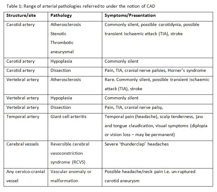
CAD itself is not a specific pathology diagnosis. Thus, there is a variation in clinical presentation which is dependent on the structure involved, the pathological mechanism and the stage at which the dysfunction presents. CAD is of relevance to clinicians due to the fact the pain is often an early presentation of many types of arterial dysfunction, Fig 1[2].

Fig 1 RK AT PhysPedia CAD.JPG

Later ischaemic manifestations of CAD, such as cranial nerve dysfunction and brain ischaemia, may be latent and develop hours, days or weeks after onset. Table 1 (below) details the range of presentations and associated clinical symptoms.

Clinically Relevant Anatomy

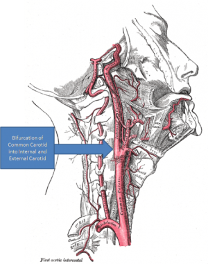
The contemporary approach to risk assessment of the cervical spine incorporates a [2]. This differs from previous paradigms by considering the complete intra and extra cranial arterial supply of the cervico-cranial region, and the wide range of associated pathologies. This includes the anterior cervical arterial system i.e. the common carotid artery (CCA) its related branches (internal and external) and pathologies, in addition to the previously considered posterior circulation, i.e. the vertebrobasilar arterial system (VBA).

473px-Gray's Anatomy with markup showing carotid artery bifurcation.png

The vertebrobasilar arterial (VBA) system and Epidemiology

Care must be taken when discussing the epidemiology. CAD is an umbrella term, and as such it is necessary to consider the epidemiology of each pathology. First, the correct population must be identified, relative to the inquiry. For example, the rate of VBI following cervical manipulation is a different matter to the prevalence of VBI in a population.  Such data may help the clinician to judge the likelihood of a patient with head/neck pain having CAD. This is a difficult question to answer with existing data as the concept is new. Nevertheless, to help judge the size of the problem, the following data can be considered.

As stroke / TIAs are a primary concern, the following data sets the scene. Ischaemic stroke is the 3rd most common cause of death in western populations[3], and in US/EU populations has a crude incidence of between 81 and 267 cases per 100,000/year[4]. Most of these events are related to atherosclerotic disease processes, with a minority related to non-atherosclerotic dissection events or mechanical phenomena. 

Historic obsession in manual therapy with the vertebral artery and dissection in particular, means that the only data available pertains to dissection of the vertebral artery following manipulation. This data is of course important but represents a very small part of a big picture. No consideration has previously been given to the other potential structures and pathologies that may result in delayed diagnosis or adverse events.

If just these data are considered, then the best estimates from the most recent studies suggest that the risk of vertebral artery dissection following manipulation is around 6:100,000, compared to a normal occurrence of around 1:100,000[5] This relates only to dissection events, There are no known data for the rate of ICA-related events following treatment due to the fact that this notion has only recently been introduced. Further, there are no known systems for collecting and collating such adverse events, so the actual rates are unknown.

A third way of framing the epidemiology is in terms of the proportion of people visiting physiotherapists who may have or develop CAD. This is the number of people with head and/neck pain who have a CAD-related pathology as the source and cause of that pain. Unlike haemorrhagic events where a headache is most often overshadowed by focal signs, ischaemic and atherosclerotic pathologies very often present with neck pain and headache as their first and most overt symptom.

Headache attributed to cranial or cervical vascular disorder is formally classed as a secondary headache by the latest International Headache Society criteria[6]. The prevalence of this, and this as a proportion of all headaches, is unknown, but very likely to be epidemiologically small. Furthermore, it should be remembered that migraineous headache itself is a known risk factor for CAD.

The epidemiology related to identifying those neuromusculoskeletal patients who may be more at risk of developing CAD is of course integrated with the above data, but even more complex to specifically identify. CAD-model reasoning would involve an understanding of the above data, together with an understanding of the societal level of vascular disease – atherosclerosis is the biggest single cause of death and morbidity in the western world – integrated with assessment of features of presentation and an individuals risk factors (see below).

Summary – the overall chance of a patient presenting with head and neck pain having a CAD-related pathology is very small. Presence of specific signs and symptoms, together with collation of known risk factors will influence this a priori probability judgment.

Pathological Process

CAD is an umbrella term covering a broad spectrum of potential pathologies. These range from pre-existing underlying anatomical anomalies, Horner’s Syndrome), blindness, stroke, or at worst, death. A spectrum of potential patho-mechanisms of ischaemia and related symptoms are covered in table 1.

Differential diagnosis

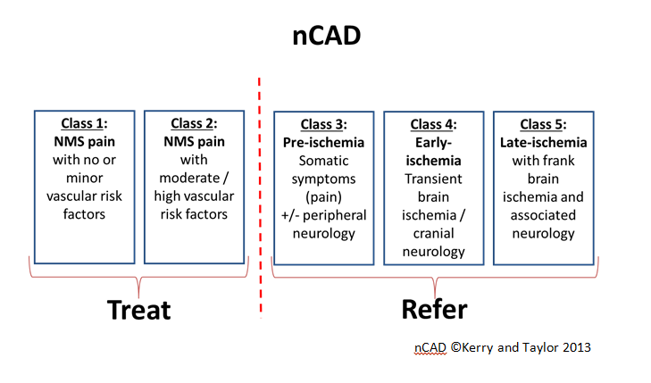
The key thing for the clinician is to be able to risk assess adequately for CAD prior to physical examination or exercise intervention. As pain may be the only initial complaint and a decision must be made as to whether this is simply a benign neuro-musculoskeletal (NMS) complaint or the pathogenesis of a neuro-vascular problem. Table 1 describes the range of possibilities.

AJT CAD Table 1.jpg

Risk factors and classification model

The following risk factors are associated with an increased risk of either internal carotid or vertebrobasilar arterial pathology and should be thoroughly assessed during the patient history:

  • Past history of trauma to cervical spine / cervical vessels
  • History of migraine-type headache
  • Hypertension
  • Hypercholesterolemia / hyperlipidemia
  • Cardiac disease, vascular disease, previous cerebrovascular accident or transient ischaemic attack
  • Diabetes mellitus
  • Blood clotting disorders / alterations in blood properties (e.g. hyperhomocysteinemia)
  • Anticoagulant therapy
  • Long-term use of steroids
  • History of smoking
  • Recent infection
  • Immediately post-partum
  • Trivial head or neck trauma
  • Absence of a plausible mechanical explanation for the patient’s symptoms.

The role of the clinician is not to aim for a precise ‘diagnosis’ of a specific condition. Rather, the CAD model facilitates the clinician in making a probability judgements on either the presence or not of any CAD-related pathology, or the likelihood of impending risk related to planned neuromusculoskeletal assessment or intervention. Figure 2[7]. provides a profiling system is designed to organise the clinicians thought processes regarding classification of patients 

.Figure 2: The Nottingham CAD (nCAD) classification

Examination

The Importance of observation throughout history

Signs and symptoms of serious pathology and contraindications / precautions to treatment may manifest during the patient history stage of assessment. This is an opportunity to observe and recognise possible red flag indicators such as gait disturbances, subtle signs of disequilibrium, upper motor neuron signs, cranial nerve dysfunction, and behaviour suggestive of upper cervical instability (e.g. anxiety, supporting head/neck) or associated ischaemia, early in the clinical encounter.

Key presenting features of CAD

It is important to consider the information below in the context of the aforementioned risk factors.

Internal carotid artery disease Vertebrobasilar artery disease Early presentation

Acute onset of pain described as “unlike any other”.

Mid-upper cervical pain, pain around ear and jaw (carotidynia),
Head pain (fronto-temporo-parietal);
Lower cranial nerve dysfunctions (VIII-XII);
 

Mid-upper cervical pain; occipital headache;
Acute onset of pain described as “unlike any other”.
Tenderness and sensitivity on the scalp
Jaw claudication (jaw pain when chewing)
Tongue claudication (tongue pain when chewing)
Late presentation

Lower cranial nerve dysfunctions (VIII-XII);

Transient retinal dysfunction (scintillating scotoma, amaurosis fugax);

Hindbrain transient ischaemic attack

(dizziness, diplopia, dysarthria,

dysphagia, drop attacks, nausea,

nystagmus, facial numbness)

Cranial nerve dysfunctions;

Ataxia, vomiting, hoarseness,

loss of short term memory, vagueness,

hypotonia/limb weakness [arm or leg],

anhidrosis [lack of facial sweating],

hearing disturbances, malaise,

Reduced visual acuity (blurred vision)

Acute visual loss (sudden blindness)
Diplopia (double vision)

Acute tinnitus (ringing in the ears)
Clinical decision-making

Some important points are made with regard to decision making for the physical examination are highlighted in the IFOMPT document.

“Based upon the evaluation and interpretation of the data from the patient history, the physical therapist needs to decide:

The above are generally normal practice for most experienced manual therapists. However, the following two items, frankly should be normal practice also, but are explicit in the document.

  • “What is the priority for these physical tests for this specific patient? What is the order of testing and to which tests should be completed at the first visit?
  • Do the physical tests need to be adapted for this specific patient?” (adapted from [8])

Management / Interventions

A patient presenting with bona fide neuromusculoskeletal condition, but with an identified risk of developing a CAD (nCAD2, e.g. smoker with hypertension and diabetes) should be treated with appropriate caution.

Physiotherapy management of a patient with a suspicion of CAD-related pathology (nCAD 3-5) requires referral for appropriate medical assessment (usually with a vascular or neurological physician).

It is essential that clinicians make appropriate triage decisions, concomitant with the urgency of the presenting condition.

Resources

Further reading on physiotherapy-specific CAD

  • Kerry R 2002 Pre-manipulative procedures for the cervical spine – new guidelines and a time for dialectics: knowledge, risks, evidence and consent. Physiotherapy 88 (7): 417-420
  • Taylor AJ and Kerry R 2005 Neck pain and headache as a result internal carotid artery dissection; Implications for clinicians. Manual Therapy 10(1): 73-77
  • Taylor AJ, Kerry R, 2005 Headache and neck pain. A sign of underlying vascular pathology? In Touch 114(4); 2-8)
  • Kerry R, Taylor AJ 2006 Masterclass: Cervical arterial dysfunction assessment and manual therapy. Manual Therapy 11(3):243-253
  • Taylor AJ, Kerry R 2008 Challenging editorial wisdom and raising the VBI debate. Manual Therapy 13(3) e5
  • Kerry R, Taylor AJ, Mitchell JM, McCarthy C, Brew J. 2008 Manual therapy and cervical arterial dysfunction – directions for the future. Journal of Manual and Manipulative Therapy 16(1): 39-48
  • Taylor AJ, Kerry R. 2008 How to recognise vascular flow problems in athletes – a clinical reasoning exercise. Sportex Medicine.
  • Kerry R, Taylor AJ. 2008 Arterial pathology and cervicocranial pain – differential diagnosis for manual therapists and medical practitioners. International Musculoskeletal Medicine 30(2): 70-77
  • Kerry R, Taylor AJ, Mitchell, JM, McCarthy C. 2008. Cervical arterial dysfunction and manual therapy: A critical literature review to inform professional practice. Manual Therapy 13(4): 278-288
  • Kerry R, Taylor AJ 2009 Cervical arterial dysfunction: knowledge and reasoning for manual physical therapists. Journal of Orthopaedic and Sports Physical Therapy 39(5):378-387
  • Taylor AJ, Kerry R 2010 A systems based approach to risk assessement of the cervical spine prior to manual therapy. Interntaional Journal of Osteopathic Medicine 13(3):85-93
  • Taylor AJ, Kerry R 2013 Vascular profiling: Should manual therapists take blood pressure? Manual Therapy. 18(4):351-353
  • Rushton A, Rivett D, Carlesso L, Flynn T, Hing W, Kerry R, International framework for examination of the cervical region for potential of cervical arterial dysfunction prior to orthopaedic manual therapy intervention, Manual Therapy 2013, doi:10.1016/j.math.2013.11.005.
  • Taylor AJ, Kerry R 2005 Vascular syndromes presenting as pain of spinal origin. Chapter 36 in: Jull G, Boyling J (eds) Grieve’s Modern Manual Therapy, 3rd edition. p517-532 Elsevier Churchill Livingstone, London
  • Kerry R, Taylor AJ 2010 Haemodynamics. Chapter 6 in: McCarthy C (ed) Combined Movement Theory. Elsevier, London

References

  1. Kerry R, Taylor AJ, Mitchell J, McCarthy C, Brew J. 2.02.1 Taylor AJ, Kerry R 2010 The Task Force on the Diagnosis and Treatment of Peripheral Artery Diseases of the European Society of Cardiology (ESC). Younan Zhang, Ann-Marie Chapman, Melanie Plested, Daniel Jackson, and Francisco Purroy. Boyle E, Côté P, Grier AR, Cassidy. Arnold M. Headache Classification Committee of the International Headache Society (IHS) The International Classification of Headache Disorders. Cephalalgia. 2018;38(1):1-211. Available from: Taylor AJ, Kerry R 2013 Vascular profiling: Should manual therapists take blood pressure? Manual Therapy. 18(4):351-353
  2. 8.08.1 Rushton A, Rivett D, Carlesso L, Flynn T, Hing W, Kerry R. International Framework for Examination of the Cervical Region for potential of Cervical Arterial Dysfunction prior to Orthopaedic Manual Therapy Intervention. International Federation of Orthopaedic Manipulative Physical Therapists (IFOMPT)fckLR2012

Further reading

  • Debette S, Leys D. Cervical-artery dissections: predisposing factors, diagnosis, and outcome. Lancet Neurol. 2009 Jul;8(7):668-78. doi: 10.1016/S1474-4422(09)70084-5. Review. PubMed PMID: 19539238.
  • Metso TM, Tatlisumak T, Debette S, Dallongeville J, Engelter ST, Lyrer PA, Thijs V, Bersano A, Abboud S, Leys D, Grond-Ginsbach C, Kloss M, Touzé E, Pezzini A, Metso AJ; CADISP group. Migraine in cervical artery dissection and ischemic stroke patients. Neurology. 2012 Apr 17;78(16):1221-8. doi: 10.1212/WNL.0b013e318251595f. Epub 2012 Apr 4. PubMed PMID: 22491867.
  • Debette S, Metso TM, Pezzini A, Engelter ST, Leys D, Lyrer P, Metso AJ, Brandt T, Kloss M, Lichy C, Hausser I, Touzé E, Markus HS, Abboud S, Caso V, Bersano A, Grau A, Altintas A, Amouyel P, Tatlisumak T, Dallongeville J, Grond-Ginsbach C; CADISP-group. CADISP-genetics: an International project searching for genetic risk factors of cervical artery dissections. Int J Stroke. 2009 Jun;4(3):224-30. doi: 10.1111/j.1747-4949.2009.00281.x. PubMed PMID: 19659826.
  • Metso TM, Debette S, Grond-Ginsbach C, Engelter ST, Leys D, Brandt T, Pezzini A, Bersano A, Kloss M, Thijs V, Lyrer PA, Tatlisumak T, Metso AJ. Age-dependent differences in cervical artery dissection. J Neurol. 2012 Oct;259(10):2202-10. doi: 10.1007/s00415-012-6485-7. Epub 2012 Apr 18. PubMed PMID: 22527225.
  • Debette S. Pathophysiology and risk factors of cervical artery dissection: what have we learnt from large hospital-based cohorts? Curr Opin Neurol. 2014 Feb;27(1):20-8. doi: 10.1097/WCO.0000000000000056. PubMed PMID: 24300790.
  • Engelter ST, Grond-Ginsbach C, Metso TM, Metso AJ, Kloss M, Debette S, Leys D, Grau A, Dallongeville J, Bodenant M, Samson Y, Caso V, Pezzini A, Bonati LH, Thijs V, Gensicke H, Martin JJ, Bersano A, Touzé E, Tatlisumak T, Lyrer PA, Brandt T; Cervical Artery Dissection and Ischemic Stroke Patients Study Group. Cervical artery dissection: trauma and other potential mechanical trigger events. Neurology. 2013 May 21;80(21):1950-7. doi: 10.1212/WNL.0b013e318293e2eb. Epub 2013 May 1. PubMed PMID: 23635964.
  • Mughal MM, Khan MK, DeMarco JK, Majid A, Shamoun F, Abela GS. Symptomatic and asymptomatic carotid artery plaque. Expert Rev Cardiovasc Ther. 2011 Oct;9(10):1315-30. doi: 10.1586/erc.11.120.
  • Kapoor S 2013 Headache Attributed to Cranial or Cervical Vascular Disorders Curr Pain Headache Rep 17:334 DOI 10.1007/s11916-013-0334-y

Deixe um comentário

O seu endereço de e-mail não será publicado. Campos obrigatórios são marcados com *

0
    0
    Suas Inscrições
    você não fez nenhuma inscriçãoRetorne ao site
    ×

    Ola! 

    Como podemos ajudar? 

    × Como posso te ajudar?