Introduction
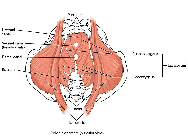
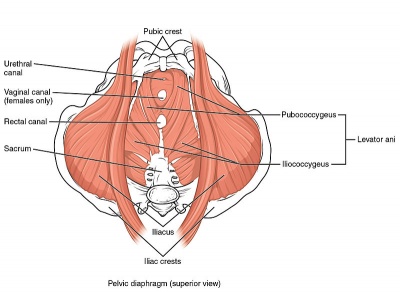
The Pelvic Floor Muscles (PFM) are found in the base of the pelvis. There are superficial muscles as well as the deep levator ani muscles. Changes in their function and strength can contribute to Pelvic Floor Dysfunction, such as urinary or faecal incontinence, pelvic organ prolapse and pelvic pain.
Pelvic Floor Muscle Function
The pelvic floor muscles provide several important functions such as pelvic organ support, bladder and bowel control and sexual function. The ability to contract the pelvic floor correctly can be difficult. A proper pelvic floor contraction incorporates both a squeeze and a lift without contraction of other muscles such as the adductors and gluts.[1]
Pelvic Floor Muscle Strength
The pelvic floor muscles need to have the ability to maintain a sustained resting tone as well as quick contractions for continence and sexual function as well as the ability to relax to allow for urination and defecation.
Evaluating the strength of the pelvic floor muscles is not as simple as assessing single maximum voluntary contraction. Factors such as power, endurance, the speed of contraction and ability to relax all need to be evaluated.
Importance of Proper Evaluation of Pelvic Floor Muscle and Function
Correct contraction of the pelvic floor cannot be adequately observed externally. In people with pelvic floor dysfunction, they often struggle to contract the pelvic floor correctly and a thorough examination will allow for an individualised treatment programme.
Assessment methods
There are various methods that can be used to evaluate the pelvic floor muscles. There is no absolute gold standard for pelvic floor muscle assessment at the moment. Examination of each patient needs to be individualised to what they are comfortable with.
Examination needs to look at the ability of the pelvic floor to contract correctly (squeeze and lift) as well as the strength and endurance of the actual contraction.[1]
External Observation
External visual observation of the perineum visualizes what happens during contraction of the pelvic floor muscles. It is usually the initial step is assessing pelvic floor muscle function.
It is not appropriate to use observation as the only assessment method as the inward movement of the skin may be created by contraction of the superficial perineal muscles and there may be no contraction of the deeper levator ani muscles.[1]
Internal Vaginal Digital Palpation
Internal digital examination of the vagina is a helpful examination tool. It is performed by inserting a finger (or fingers) into the vaginal cavity.
Pelvic floor muscle contraction can be felt and the therapist is looking for both a squeeze and lift.
When doing an internal vaginal palpation various aspects of pelvic floor muscle strength need to be examined.
Laycock[2] developed the PERFECT Scheme which is a method of examination of the pelvic floor muscles that looks at
- Power – Modified Oxford Scale)
- Endurance – How long can they hold a maximal voluntary contraction (up to 10sec)
- Repetitions – How many maximal voluntary contractions they hold with a rest between them, up to 10 reps (eg 10 repetitions of a 10-second hold)
- Fast- The number of 1 second maximal voluntary contractions they can perform in a row (up to 10)
- Every Contraction Timed – a reminder to time every contraction
An internal vaginal examination has good intra-rater reliability but only fair intrer-rater reliability [3]. This means that follow up internal examinations by the same practitioner can provide useful information in clinical practice. However findings between different examiners may not be as accurate
The advantage of an internal vaginal examination is that as well as the muscle strength, anatomical changes, the symmetry of a contraction, muscle tone as well as any painful areas can also be examined [3][4]
Ultrasound [5][4]
Real-time trans-abdominal and trans-perineal ultrasound can both be used to assess pelvic floor contractions. Both have shown to be reliable in assessing movement of pelvic structures during contraction[5].
Transabdominal ultrasound is a non-invasive, valid and reliable tool that can be used to assess pelvic floor muscle contraction[4]. By placing the ultrasound probe supra-pubically the examiner can evaluate both the squeeze and lift component of the contraction[4]. The strength of muscle contractions cannot be accurately assessed on ultrasound but the ability to contract and endurance can be evaluated accurately.
Real-time ultrasound is helpful as it provides a good visual feedback tool for patients to use as a means of contracting their pelvic floor muscles correctly. Transabdominal ultrasound is also non-invasive so can be used in patients who are not comfortable with an internal examination or where an internal examination is contra-indicated eg children.
MRI
MRI can be used to evaluate for pelvic floor dysfunction as well as the ability of the pelvic floor to lift while contracting. Clinically it is very expensive and is not used widely for standard PFM assessment.
EMG
Surface Electromyography can be used to evaluate the pelvic floor muscles,
PFM activation recorded using transperineal sEMG is only weakly correlated with PFM strength. Results from perineal sEMG should not be interpreted in the context of reporting PFM strength.[3]
Manometers and Dynamometers
Manometry and dynamometry are reliable tools that can measure a pelvic floor muscle contraction and may be superior to digital palpation as an objective measure of strength.[3]
Conclusion
There are a variety of ways to assess PFM function and strength. Real-time ultrasound and internal digital palpation are probably the most widely used in clinical practice as they are the most cost-effective and efficient means of examination. Manometry and Dynamometry may provide a more accurate measurement of strength.
When assessing PFM function and strength it is important to correlate multiple examination findings to ensure accurate evaluation.


