Description
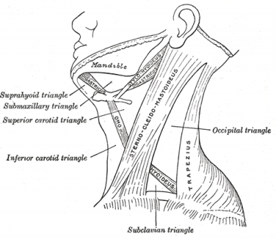
Sternocleidomastoid (SCM) (synonym musculus sternocleidomastoideus) is a paired superficial muscle in the anterior portion of the neck. The sternocleidomastoid muscle (SCM) is an important landmark in the neck which divides it into an anterior and a posterior triangle. This muscle binds the skull to the sternum and clavicle.[2] It protects the vertical neurovascular bundle of neck, branches of cervical plexus, deep cervical lymph nodes and soft tissues of neck from damage [2]
Origin
It arises by two heads:
- a medial rounded and tendinous sternal head (SH)
- a lateral fleshy clavicular head (CH). [3]
They arise from the anterolateral surface of the manubrium sterni and the medial third of the superior surface of the clavicle, respectively. The thickness of the CH is variable. The two heads are separated by a triangular surface depression, the lesser supraclavicular fossa. As they ascend, the CH spirals behind the SH and blends with its deep surface below the middle of the neck, forming a thick rounded belly. [2]
Insertion
Lateral surface of the mastoid process through a strong tendon, and to the lateral half of superior nucheal line through an aponeurosis.
Nerve Supply
Spinal accessory nerve (XI), with sensory supply from C2 & C3 (for proprioception)
Blood Supply
Sternocleidomastoid branch of the Occipital artery
Action
Draws the mastoid process down toward the same side which causes the chin to turn up toward the opposite side; acting together, the muscles of the two sides flex the neck
Function
Rotation of the head to the opposite side or obliquely rotate the head. It also flexes the neck. When acting together it flexes the neck and extends the head. When acting alone it rotates to the opposite side (contralaterally) and slightly (laterally) flexes to the same side. It also acts as an accessory muscle of inspiration.
Morphological variations of the SCM
- The SCM is a unique muscle, in terms of variations at its origin.4,5,6 Also, it has a variable innervations arrangement, the “ classical anastomotic pattern “ being observed in 50 % of the cases.These anatomical details have a pivotal role in the planning of pedicle muscle flaps in reconstructive surgeries. [5]
- In many animals, the cleidomastoid belly is distinctly separate from the sternomastoid belly. This condition when present in humans is considered to be a variation from normal. The two separate sternomastoid and cleidomastoid bellies further subdivide the anterior triangle into a supernumerary triangle. This extra triangle can also be considered as an extended lesser supraclavicular fossa which normally separates the sternal and clavicular heads of origin of SCM. The occurrence of such a variation can be explained by fusion failure or abnormal mesodermal splitting during development. In this regard we may refer to Sinohara’s law of fusion which states that a muscle supplied by two different nerves is formed by fusion of two separate muscle masses. [5]
- Some studies have indicated a supernumerary cleido-occipital muscle more or less separate from the sterno-cleido-mastoid muscle. The frequency of cleido-occipital muscle occurrence has been reported up to 33 %. [5]
- Occasionally, the SCM fuses with the trapezius, leaving no posterior triangle. Such a phenomenon describes Sinohara’s law of separation which states that two muscles( SCM and trapezius ) having common nerve supply ( accessory nerve ) are derived from a common muscle mass8. Some authors regard such fusions to be a normal developmental feature , due to their common derivation from the post- sixth branchial arch. [5]
- There are reports of a broad clavicular head splitting into multiple small muscular slips. The number of these extra clavicular slips may vary and such occurrence may be unilateral or bilateral. They cause formation of supernumerary lesser supraclavicular fosse. Developmentally, these additional muscle slips indicate abnormal mesodermal splitting in posterior sixth branchial arch. They may not cause any functional advantage or disadvantage in neck movement but might be physically interfering during invasive procedures. However, they can be effectively utilized for muscle flap harvests.
- There are also cases presenting with extra sternal and clavicular heads of origin in SCM.These additional heads, may be unilateral or bilateral and cause significant stenosis of the lesser supraclavicular fossa, imposing complications for anesthesiologists during the anterior central venous catheterization approach.
- Benign fibrosis, hypoplasia or aplasia of SCM is the most common cause of congenital torticolis. Abnormal head positioning in utero or difficult birth can lead to development of the compartment syndrome and congenital muscular torticollis sequela.Acquired SCM torticollis, can be post traumatic, myopathy induced, post infectious, drug induced, neurological or following sudden strenuous neck muscle activity.
- Studies report that morphometric and cross-sectional area a-symmetry between SCM of two sides result from unequal growth in utero and play an important role in the genesis of tension type headache. [5]
- About a dozen cases have reported complete unilateral absence of the muscle. Such cases represent the developmental defect of muscular agenesis and are diagnosed by Ultrasound or Magnetic Resonance Imaging scans. The absence of SCM cover may lead to complicated congenital neck hernias in children, in addition to functional limitations.
- A coexisting unilateral absence of SCM with the ipsilateral absent trapezius is an extremely rare variation and till date, only about three such reports are present in literature .Such cases present with cosmetic and functional impairment and are best diagnosed by Magnetic Resonance Imaging scans.
- Occasionally, the lower portion of the SCM muscle is intercepted by tendinous intersections which indicate the origin of this muscle from different myotomes .The organizational pattern of the SCM can be arranged into five distinct topographical parts, namely the superficial sternomastoid, profound sternomastoid, sterno occipital, cleidomastoid and cleidooccipital parts which are arranged in superficial and deep layers. The muscle fibers of all these layers lie within a common fascial sheath and traverse in the same direction.Knowledge of this layered arrangement and the changes in cases of muscle variations is helpful during muscle flap harvesting procedures.
References
- ↑ T Hasan. Variations Of The Sternocleidomastoid Muscle: A Literature Review. The Internet Journal of Human Anatomy, 2010
- ↑ 2.02.12.2 Kaur D et al. Six heads of origin of sternocleidomastoid muscle: a rare case. Internet Journal of Medical Update 2013; 8(2):62-64
- ↑ Gray, Henry. Anatomy of the Human Body. Philadelphia: Lea and Febiger, 1918; Bartleby.com, 2000
- ↑ KenHub. Sternocleidomastoid. Available from: ↑ 5.05.15.25.35.4 T Hasan. Variations Of The Sternocleidomastoid Muscle: A Literature Review. The Internet Journal of Human Anatomy 2010


