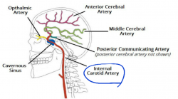
It is terminal branch of the common carotid artery, it is larger than the other terminal branch (the external carotid artery).[1]
There are seven segments according to Bouthillier classification:
- C1/ Cervical segmen
- C2/ Petros (horizontal) segment
- C3/ Lacerum segment
- C4/ Cavernous segment
- C5/ Clinoid segment
- C6/ Ophthalmic (Supra clinoid) segment
- C7/ Communicating segment
The odd numbered segments usually have no branches except for the terminal segment C7 while it has four branches, whereas each even numbered segment (C2, C4 and C6) has two branches:
| Segment | branches |
| C1 | non |
| C2 |
1.caroticitympanic artery 2.vidian artery |
| C3 | non |
| C4 |
1.Meningohypophyseal trunk 2. Inferiolateral trunk |
| C5 | non |
| C6 |
1. Ophthalmic artery 2. Superior hypophyseal artery |
| C7 |
1. Posterior communicating artery 2. Anterior chorodal artery 3. Anterior cerebral artery 4. Middle cerebral artery |
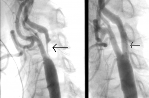
Internal carotid artery stenosis:
It is an important cause of ipsilateral stroke. The natural history of the disease is related t the presence or absence of ipsilateral hemispheric symptoms and the severity of stenosis.[2]
Complete occlusion of the internal carotid artery (ICA):
It is an important cause of cerebrovascular disease. A nerve_symptomatic occlusion increases future risk of strokes.
Ultrasonography, magnetic resonance imaging and contrast angiography are useful diagnostic tests and functional imaging of the brain helps to understand haemodynamic factors involved in the pathophysiology of brain ischaemia.[3]
- ↑ 1.01.11.2 Internal carotid artery, ↑ Lanzino G, TallaritaT,Rabinstein AA, Internal carotid artery stenosis: natural history and management, SeminNeurol, 2010 Nov;30(5):518:27
- ↑ BhomrajThanvi and Tom Robinson, complete occlusion of extracranial internal carotid artery: clinical features, pathophysiology, diagnosis and management, Postgrad Med J. 2007 Feb; 83(976): 95-99.


