Definition/Description
Chondromalacia patellae (CMP) is referred to as anterior knee pain due to the physical and biomechanical changes [1]. The articular cartilage of the posterior surface of the patella is going though degenerative changes [2] which manifests as a softening, swelling, fraying, and erosion of the hyaline cartilage underlying the patella and sclerosis of underlying bone. [3]
Chondromalacia patellae is one of the most frequently encountered causes of anterior knee pain among young people. It’s the number one cause in the United States with an incidence as high as one in four people.[4] The word chondromalacia is derived from the Greek words chrondros, meaning cartilage and malakia, meaning softening. Hence chondromalacia patellae is a softening of the articular cartilage on the posterior surface of the patella which may eventually lead to fibrillation, fissuring and erosion.[5]
The differential diagnosis of chondromalacia include patellofemoral pain syndrome and patellar tendinopathy. Chondromalacia is are not considered to be under the umbrella term of PFPS.[6][7][8] The pathophysiology is thought to be different and therefore there is alternative treatment.[6][8]
Clinically Relevant Anatomy
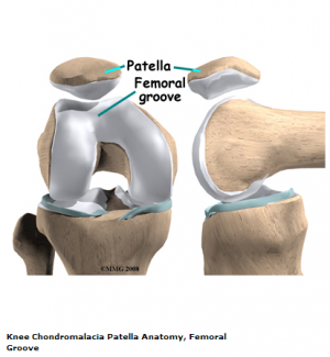
The knee comprises of 4 major bones: the femur, tibia, fibula and the patella. The patella articulates with the femur at the trochlear groove. [9] Articular cartilage on the underside of the patella allows the patella to glide over the femoral groove, necessary for efficient motion at the knee joint. [10] Excess and persistent turning forces on the lateral side of the knee can have a negative effect on the nutrition of the articular cartilage and more specifically in the medial and central area of the patella, where degenerative change will occur more readily. [11]
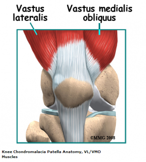
The quadriceps insert into the patella via the quadriceps tendon and are divided into four separate muscles: rectus femoris (RF), vastus lateralis (VL), vastus intermedius (VI) and vastus medialis (VM). The VM has oblique fibres which are referred to the vastus medialis obliques (VMO)[12]
These muscles are active stabilisers during knee extension, especially the VL (on the lateral side) and the VMO (on the medial side). The VMO is active during knee extension, but does not extend the knee. Its function is to keep the patella centred in the trochlea. This muscle is the only active stabiliser on the medial aspect, so it’s functional timing and amount of activity is critical to patellofemoral movement, the smallest change having significant effects on the position of the patella.
Not only do the quadriceps influence patella position, but also the passive structures of the knee. These passive structures are more extensive and stronger on the lateral side than they are on the medial side, with most of the lateral retinaculum arising from the iliotibial band (ITB). If the ITB is under excessive tension, excessive lateral tracking and/or lateral patellar tilt can occur. This is can be as a result of the tensor fasciae lata being tight, as the ITB itself is a non contractile structure. [11].
Other significant anatomical structures:
- Femoral anteversion [13] or medial torsion of the femur is a condition which changes the alignment of the bones at the knee. This may lead to overuse injuries of the knee due to malalignment of the femur in relation to the patella and tibia. [14]
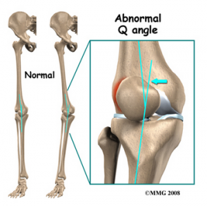
- The Q-angle: or quadriceps angle is the geometric relationship between the pelvis, the tibia, the patella and the femur [14] [15] and is defined as the angle between the first line from the anterior superior iliac spine to the centre of the patella and the second line from the centre of the patella to the tibial tuberosity [16].
If there is an increased adduction and/or internal rotation of the hip, the Q-angle will increase, which increases the relative valgus of the lower extremity as well. This higher Q-angle and valgus will increase the contact pressure on the lateral side of the patellofemoral joint (which is also increased by external rotation of the tibia) [17]
Epidemiology /Etiology
The etiology of CMP is poorly understood, although it is believed that the causes of chondromalacia are injury, generalised constitutional disturbance and patellofemoral contact [18], or as a result of trauma to the chondrocytes in the articular cartilage (leading to proteolytic enzymatic digestion of the superficial matrix). It may also be caused by instability or maltracking of the patella which softens the articular cartilage. [19] Chondromalacia patella is usually described as an overload injury, caused by malalignment of the femur to the patella and the tibia. [20]
Main reasons for patellar malalignment;
- Q-angle: An abnormality of the Q-angle is one of the most significant factors of patellar malalignment. A normal Q-angle is 14° for men and 17° for women. An increase can result in an increased lateral pull on the patella.
- Muscular tightness of:
> Rectus femoris: affects patellar movement during flexion of the knee.
> Tensa Fascia late; affects the influence of the ITB
> Hamstrings: during running tight hamstrings increase knee flexion which results in increased ankle dorsiflexion. This causes compensatory pronation in the talocrural joint.
> Gastrocnemius: tightness will result in compensatory pronation in the subtalar joint.
- Excessive pronation: prolonged pronation of the subtalar joint is caused by internal rotation of the leg. This internal rotation will result in malalignment of the patella.
- Patella alta: this is a condition where the patella is positioned in an abnormally superior position. It is present when the length of the patellar tendon is 20% greater than the height of the patella.
- Vastus medialis insufficiency: the function of the vastus medialis is to realign the patella during knee extension. If the strength of VM is insufficient this will cause a lateral drift of the patella.[21]
Muscular balance between the VL and VM is important. Where VM is weaker the patella is pulled too far laterally which can cause increased contact with the condylus lateralis, leading to degenerative disease.[22]
Degenerative changes of the articular cartilage can be caused by [23]:
- Trauma: instability caused by a previous trauma or overuse during recovery
- Repetitive micro trauma and inflammatory conditions
- Postural distortion: causes malposition or dislocation of the patella in the trochlear groove
Hip positioning and strength are linked to the prevalence of patellofemoral pain syndrome. Therefore, hip strengthening and stability exercises may be useful in the treatment program of patellofemoral pain syndrome.[17]
Some authors use the term “patellar pain syndrome” instead of “chondromalacia” in order to describe “anterior knee pain”. [24]
Stages of the disease
In the early stages, chondromalacia shows areas of high sensitivity on fluid sequences. This can be associated with the increased thickness of the cartilage and may also cause oedema. In the latter stages, there will be a more irregular surface with focal thinning that can expand to and expose the subchondral bone. [25]
Chondromalacia patella is graded based on the basis of arthroscopic findings, the depth of cartilage thinning and associated subchondral bone changes. Moderate to severe stages can be seen on MRI. [26]
- Stage 1: softening and swelling of the articular cartilage due to broken vertical collagenous fibres. The cartilage is spongy on arthroscopy.
- Stage 2: blister formation in the articular cartilage due to the separation of the superficial from the deep cartilaginous layers. Cartilaginous fissures affecting less than 1,3 cm² in area with no extension to the subchondral bone.
- Stage 3: fissures ulceration, fragmentation, and fibrillation of cartilage extending to the subchondral bone but affecting less than 50% of the patellar articular surface.
- Stage 4: crater formation and eburnation of the exposed subchondral bone more than 50% of the patellar articular surface exposed, with sclerosis and erosions of the subchondral bone. Osteophyte formation also occurs at this stage.
Articular cartilage does not have any nerve endings, so CMP should not be considered as a true source of anterior knee pain, rather, it is a pathological or surgical finding that represents areas of articular cartilage trauma or divergent loading. [10]Kok et al showed that there is significant association between subcutaneous knee fat thickness with the presence and severity of chondromalacia patellae. This could explain why women suffer more from the condition chondromalacia than men. [27]
Characteristics/Clinical Presentation
There are important distinguishing features between chondromalacia patellae and Osteoarthritis. CMP affects just one side of the joint, the convex patellar side, [28] with excised patellas show localised softening and degeneration of the articular cartilage. [29] The main symptom of chondromalacia patellae is anterior knee pain,[18] which is exacerbated by common daily activities that load the patellofemoral joint, such as running, stair climbing, squatting, kneeling [1], or changing from a sitting to a standing position [30]. The pain often causes disability affecting the short term participation of daily and physical activities.[31] Other symptoms are tenderness on palpating under the medial or lateral border of the patella, [32] crepitation (felt with motion), [33]; minor swelling, [32] a weak vastus medialis muscle and a high Q-angle. [34] Vastus medialis is functionally divided into two components: the vastus medialis longus (VML) and the vastus medialis obliquus (VMO). The VML extends the knee, with the rest of the quadriceps muscle. The VMO does not extend the knee, but is active throughout knee extension. This component assists in keeping the patella centred in the femoral trochlea. [11]
This condition can cause a deficit in quadricep strength, therefore, building and/or maintaining quadriceps strength is essential.[1] A significant number of individuals are asymptomatic, but crepitation in flexion or extension is often present. [35]Chondromalacia is common in adolescents and females with idiopathic chondromalacia usually seen in young children and adolescents and the degenerative condition is most common in the middle aged and older population. [25]
Differential Diagnosis
Diagnostic Procedures
Since its first description by Budinger in 1906, chondromalacia patella has been of significant clinical interest because diagnosis is often difficult. The chief reason for this is that the aetiology is often unknown and the correlation between the articular cartilage changes and the clinical system is poor. Patients affected by chondromalacia patella are young, between 15 and 35 years old, and many are highly active and are often considerably disabled by the symptoms of aching behind the patella, recurrent effusion of the knee, knee instability and crepitus.[36]
The primary diagnostic approach for chondromalacia patellae is radiography with added arthrography. Pinhole scintigraphy, part of arthrography, is also used to diagnose the condition. [37] MRI is an effective, non-invasive method with the ability to increase the sensitivity and specificity of the diagnosis.[38]
Outcome Measures
There are various measures: [39][40]
- Anterior Knee Pain Scale: a 13 item questionnaire with categories related to various levels of current knee function.
- Visual analog scale
- The five KOOS subscales: a scale about patients’ experience over time with knee conditions. It consists of five subscales: Pain, other Symptoms, Function in daily living, Function in sport and recreation and knee related Quality of life.
Examination
Examination of the knee is 4 fold: observation, mobility, feel, X-ray.[41]
Observation: joint appearance is usually normal, but there may be a slight effusion.
Mobility: passive movements are usually full and painless, but repeated extension of the knee from flexion will produce pain and a grating feeling underneath the patella, especially if the articular surfaces are compressed together.
Feel: Pain and crepitus will be felt if the patella is compressed against the femur, either vertically or horizontally, with the knee in full extension. By displacing the patella medially or laterally, the patellar margins and their articular surfaces may be felt. Tenderness of one or other margin may be elicited and more frequently the felt medially. Resisting a static quadriceps contraction, will generally produce a sharp pain under the patella. This may be apparent in both knees, but more severe on the affected side.
X-ray: an AP view of the patellofemoral joint is needed to detect any radiological change. In all but the most advanced cases, there is no convincing radiological change. In the latter stages, patellofemoral joint space narrows and osteoarthritic changes begin to appear.
Tests
The patient’s posture can be an initial clue as well as any observed asymmetries, such as; limb alignment in standing, internal femoral rotation, anterior or posterior pelvic tilt, hyperextended or ‘locked back’ knees, genu varum or valgum and abnormal pronation of the foot. Gait pattern may also be affected. [12]
Mobility and range of motion (ROM) of the joint are tested, which can be limited. if bursitis is present, passive flexion or active extension will be painful. Loss of power in the affected leg may also be present on isometric testing. There are specific tests for anterior knee pain syndrome: [33]
It is possible to diagnose incorrectly and these tests may aid in determining chondromalacia, but other possible conditions also need to be excluded.
Medical Management
Exercise and education are two important aspects of a treatment programme. Education helps the patient to understand the condition and how they should deal with it for optimal recovery. Exercise focus is on stretching and strengthening appropriate structures, such as: hamstring, quadriceps and gastrocnemius length and strength of the gluteal muscles.[43] Fire needling and acupuncture may also relieve clinical symptoms of chondromalacia patellae and recovers the biodynamical structure of patellae. [44]
If conservative measures fail, there are a number of possible surgical procedures. [26]
Chondrectomy: also known as shaving. This treatment includes shaving down the damaged cartilage to the non damaged cartilage underneath. The success of this treatment depends on the severity of the cartilage damage.
Drilling is also a method that is frequently used to heal damaged cartilage. However, this procedure has not so far been proven to be effective. More localised degeneration might respond better to drilling small holes through the damaged cartilage. This facilitates the growth of the healthy tissue through the holes from the layers underneath.
Full patellectomy: This is the most severe surgical treatment. This method is only used when no other procedures were helpful, but a significant consequence is that the quadriceps will become weak.
Two other treatments that may be successful: [23]
- Replacement of the damaged cartilage : The damaged cartilage is replaced by a polyethylene cap prosthesis. Early results have been good, but eventual wearing of the opposing articular surface is inevitable.
- Autologous chondrocyte transplantation under a tibial periosteal patch. [23]
Simply removing the cartilage is not a cure for chondromalacia patellae. The biomechanical deficits need addressing and there are various procedures to aid in managing this.
- Tightening of the medial capsule (MC): If the MC is lax, it can be tightened by pulling the patella back into its correct alignment.
- Lateral release: A very tight lateral capsule will pull the patella laterally. Release of the lateral patellar retinaculum allows the patella to track correctly into the femoral groove.
- Medial shift of the tibial tubercle: Moving the insertion of the quadriceps tendon medially at the tibial tubercle, allows the quadriceps to pull the patella more directly. It also decreases the amount of wear on the underside of the patellar.
- Partial removal of the patella
Although there is no overall agreement for the treatment of chondromalacia, the general consensus is that the best treatment is a non-surgical one.[45]
Physical Therapy Management
Exercise Program
Conservative treatment of chondromalacia patellae is both physical and highly advised. Short-wave diathermy can help to relieve pain and to increase the blood supply to the area, improving nutrition supply to the articular cartilage. Care must be taken when planning an exercise programme. (Level of Evidence 2B)[43] Conservative therapeutic interventions include the following: [52]
- Isometric quadriceps strengthening and stretching exercises (Level of Evidence 2B )[1] Restoration of adequate quadriceps strength and function is an essential factor in achieving good recovery.The most effective exercises are isometric and isotonic in the inner range. Isotonic exercises through a full range of motion will only lead to increased pain and even joint effusion.[43] Stretching of the vastus lateralis and strengthening of the vastus medialis is often recommended, but they are difficult to isolate due to shared innervation and insertion. (Level of Evidence 1A)[12][22]It has shown that closed kinematic chain exercices can improve patellofemoral joint performance by increasing quadriceps muscle strenght and patellar alingment correction. (Level of Evidence 1A)[46]
- Hamstring stretching exercises
- Temporary modification of activity
- Patellar taping
- Foot orthoses
- NSAIDS
- Hip strength and stability training, as hip positioning and strength has a significant influence on anterior knee pain.
- Hip abductor strengthening as an increased hip adduction angle is associated with weakened hip abductors. (Level of Evidence 1A)[47]
- Patellar realingment brace[40]
Not only is strengthening important, but stretching should also be part of the programme. (Level of Evidence 1A)[10] It ha been shown that patients with patellofemoral pain syndrome have shorter and less flexible hamstrings than asymptomatic individuals.. Although stretching can improve flexibility and knee function, it doesn’t necessarily directly improve pain. (Level of Evidence 4)[48]
Another form of therapy is warm needling. In combination with rehabilitation exercises it has a prolonged pain relieving effect than in warm needling in combination with medication. (Level of Evidence 1A)[49]
Ice medication
Ice may be useful for reducing pain in an acute flare up, but not as a long term treatment protocol.[47] NSAIDS may also be of benefit in the short term to relieve pain so that knee function and mobility is normalised and an exercise programme can begin.
Taping and braces
Taping the patella to influence its movement may provide some short term relief, but the evidence is varied. A commonly used technique is ‘McConnell taping or kinesio taping. (Level of Evidence 2B)[50][51]
Supporting the patella and knee joint by bracing is a further way to reduce pain and symptoms, but it will also alter patella tracking and reduce active function of the quadriceps. Bracing may be useful in the short term to offer patients some support and pain relief to help them avoid antalgic movements and normalise gait as much as possible. Bracing can also be used for patients pre- and postoperatively, but a brace should allow variation in medial pull on the patellar and pressure. (Level of Evidence 1A )[24] Wearing a patellar realingment brace and following physical therapy has a synergistic effect on patients with chondromalacia patellae.[40]
Foot Orthoses
Foot orthoses are another option for pain relief, but only in cases where a lower limb mechanics is deemed to be contributing to the knee pain, which may be due to: poor pronation control, excessive lower limb internal rotation during weight bearing and an increased Q-angle. (Level of Evidence 2B)[31][24]
Foam roller
Using a foam roller cab be useful for relieving tight musculature and reducing pressure over the patella. .[52]
References
- ↑ 1.01.11.21.3 Lee Herrington and Abdullah Al-Sherhi, A Controlled Trial of Weight-Bearing Versus Non–Weight-Bearing Exercises for Patellofemoral Pain, journal of orthopaedic sports physical therapy, 2007, 37(4), 155-160
Level of Evidence: 2B
- ↑ ↑ Gagliardi et al., Detection and Staging of Chondromalacia Patellae: Relative Efficacies of Conventional MR Imaging, MR Arthrography, and CT Arthrography, ARJ, 1994, 163, 629-636
- ↑ Laprade J, Culham E, Brouwer B (1998) Comparison of five isometric exercises in the recruitment of the vastus medialis oblique in persons with and without patellofemoral pain syndrome. J Orthop Sports Phys Ther 27: 197–204
- ↑ Helen M. Gordon : CHONDROMALACIA PATELLAE1 1Delivered at the XV Biennial Congress of the Australian Physiotherapy Association, Hobart, February 1977. Australian Journal of Physiotherapy, Volume 23, Issue 3, September 1977, Pages 103-106
- ↑ 6.06.1 Wiles P, Andrews PS, Devas MB. Chondromalacia of the patella. Bone & Joint Journal. 1956 Feb 1;38(1):95-113.
- ↑ Blazer K. Diagnosis and treatment of patellofemoral pain syndrome in the female adolescent. Physician Assistant. 2003 Sep 1;27(9):23-30.
- ↑ 8.08.1 Fernández-Cuadros ME, Albaladejo-Florín MJ, Algarra-López R, Pérez-Moro OS. Efficiency of Platelet-rich Plasma (PRP) Compared to Ozone Infiltrations on Patellofemoral Pain Syndrome and Chondromalacia: A Non-Randomized Parallel Controlled Trial. Diversity & Equality in Health and Care. 2017 Aug 4;14(4).
- ↑ ↑ 10.010.110.2 ANDERSON M. K. ,Fundamentals of Sports Injury Management, second edition, Lippincott Williams & Wilkins, 2003, p. 208
(levels of evidence: 5) - ↑ 11.011.111.2 BEETON K. S., Manual Therapy Masterclasses, The Peripheral Joints, Churchill Livingstone, 2003, p.50-51 fckLR(Levels of Evidence: 5E)
- ↑ 12.012.112.2 Florence Peterson Kendall et al., Spieren : tests en functies, Bohn Stafleu van Loghum, Nederland, 469p (383)
(Level of Evidence: 5) - ↑ NYLAND J et al., Femoral anteversion influences vastus medialis and gluteus medius EMG amplitude: composite hip abductor EMG amplitude ratios during isometric combined hip abduction-external rotation, Elsevier, vol. 14, issue 2, April 2004, p. 255-261. (Levels of Evidence: 2C)
- ↑ 14.014.1 MILNER C. E., Functional Anatomy For Sport And Exercise: Quick Reference, Routledge, 2008, p. 58-60 fckLR(Levels of evidence: 5E)
- ↑ SINGH V., Clinical And Surgical Anatomy, second edition, Elsevier, 2007, p. 228- 230. fckLR(Levels of Evidence: 2C)
- ↑ ASSLEN M. et al., The Q-angle and its Effect on Active Joint Kinematics – a Simulation Study, Biomed Tech 2013; 58 (suppl 1). (Levels of Evidence: 3B)
- ↑ 17.017.1 Erik P. Meira, Jason Brumitt. “Influence of the Hip on Patients With Patellofemoral Pain Syndrome: A Systematic Review.” Sports Health: A Multidisciplinary Approach, September/October 2011; vol. 3, 5: pp. 455-465
- ↑ 18.018.1 Iraj Salehi, Shabnam Khazaeli, Parta Hatami, Mahdi Malekpour, Bone density in patients with chondromalacia patella, Springer-Verlag, 2009
- ↑ MACMULL S., The role of autologous chondrocyte implantation in the treatment of symptomatic chondromalacia patellae, International orthopaedics, Jul 2012, 36(7), 1371-1377. (Levels of Evidence: 1B)
- ↑ BARTLETT R., Encyclopedia of International Sports Studies, Routledge, 2010, p. 90. (Levels of Evidence: 5F)
- ↑ Jenny McConnell. “The management of chondromalacia patellae: a long term solution.” Australian Journal of Physiotherapy, volume 32, issue 4, 1986, pages 215-223
- ↑ 22.022.1 ↑ 23.023.123.2 LOGAN A. L., The Knee Clinical Applications, Aspen Publishers, 1994, p. 131.
- ↑ 24.024.124.2 MANSKE R. C., Postsurgical Orthopedic Sports Rehabilitation: Knee & Shoulder, 2006, Mosby Elsevier, p. 446, 451.
(Level of Evidence: 5) - ↑ 25.025.1 WESSELY M., YOUNG M., Essential Musculoskeletal MRI: A Primer for the Clinician, Churchill Livingstone Elsevier, 2011, p. 115. (Levels of Evidence: 5E
- ↑ 26.026.1 MUNK P. L., RYAN A. G., Teaching Atlas of Musculoskeletal Imaging, Thieme, 2008, p. 68-70. (Levels of Evidence: 5E)
- ↑ KOK HK., Correlation between subcutaneous knee fat thickness and chondromalacia patellae on magnetic resonance imaging of the knee, Canadian Association of Radiologists journal, Aug 2013, 64(3), 182-186. (Levels of Evidence: 2B)
- ↑ ELLIS H., FRENCH H., KINIRONS M. T., French’s Index of differential diagnosis, 14th edition, Hodder Arnold Publishers, 2005. (Levels of Evidence: E)
- ↑ ANDERSON J. R., Muir’s Textbook of Pathology, 12th edition, Lippincott Williams Wilkins, 1988 (Levels of Evidence: C)
- ↑ MOECKEL E., NOORI M., Textbook of Pediatric Osteopathy, Elsevier Health Sciences 2008, p. 338. (Levels of Evidence: D)
- ↑ 31.031.1 Bill Vicenzino, Natalie Collins, Kay Crossley, Elaine Beller, Ross Darnell and Thomas McPoil, fckLRFoot orthoses and physiotherapy in the treatment of patellofemoralfckLRpain syndrome: A randomised clinical trial, BioMed Central, 2008
Level of Evidence: 1b
- ↑ 32.032.1 SHULTZ S. J., HOUGLUM P. A., PERRIN D. H., Examination of Musculoskeletal injuries, third edition, Human Kinetics, 2010, p. 453. (Levels of Evidence: E)
- ↑ 33.033.1 DEGOWIN R. L., DEGOWIN E. L., DeGowin & DeGowin’s Diagnostic Examination, 6th edition, McGraw Hill, 1994, p. 735. (Levels of Evidence: B)
- ↑ EBNEZAR J., Textbook of Orthopedics¸ 4th edition, JP Medical Ltd, 2010, p. 426-427. (Levels of Evidence: E)
- ↑ MURRAY R. O., JACOBSON H. G., The Radiology of Skeletal Disorders: exercises in diagnosis, second edition, Churchill Livingstone, 1990, p. 306-307. (Levels of Evidence: E)
- ↑ George Bentley, Ian J. Lesly and David Fischer. “Effect of aspirin treatment on chondromalacia patella” Annals of the rheumatic diseases, 1981, 40, p37-41.
- ↑ Bahk YW, Park YH, Chung SK, Kim SH, Shinn KS. “Pinhole scintigraphic sign of chondromalacia patellae in older subjects: a prospective assessment with differential diagnosis.” Journal of Nuclear Medicine : Official Publication, Society of Nuclear Medicine 1994, 35(5):855-862
- ↑ Kim, H. J., Lee, S. H., Kang, C. H., Ryu, J. A., Shin, M. J., Cho, K. J., & Cho, W. S. (2011). Evaluation of the chondromalacia patella using a microscopy coil: comparison of the two-dimensional fast spin echo techniques and the threedimensional fast field echo techniques. Korean J Radiol, 12(1), 78-88
- ↑ Crossley, Kay M., et al. “Analysis of outcome measures for persons with patellofemoral pain: which are reliable and valid?.” Archives of physical medicine and rehabilitation 85.5 (2004): 815-822.
- ↑ 40.040.140.2 Petersen, Wolf, et al. “Evaluating the potential synergistic benefit of a realignment brace on patients receiving exercise therapy for patellofemoral pain syndrome: a randomized clinical trial.” Archives of orthopaedic and trauma surgery (2016): 1-8. (level of evidence: 1b)
- ↑ Helen M. Gordon : CHONDROMALACIA PATELLAE1 1Delivered at the XV Biennial Congress of the Australian Physiotherapy Association, Hobart, February 1977. Australian Journal of Physiotherapy, Volume 23, Issue 3, September 1977, Pages 103-106
- ↑ Laprade J, Culham E, Brouwer B (1998) Comparison of five isometric exercises in the recruitment of the vastus medialis oblique in persons with and without patellofemoral pain syndrome. J Orthop Sports Phys Ther 27: 197–204
- ↑ 43.043.143.2 CLARK, D. I., N. DOWNING, J. MITCHELL, L. COULSON, E. P. SYZPRYT, and M. DOHERTY. Physiotherapy for anterior knee pain: a randomised controlled trial. Ann. Rheum. Dis. 59:700–704, 2000.
Level of Evidence: 1b
- ↑ Huang J, Li L, Lou BD, Tan CJ, Liu Z, Ye Y, Huang A, Li X, Zhang W. [Efficacy observation on chrondromalacia patellae treated with fire needling technique at high stress points] Zhongguo Zhen Jiu. 2014 Jun;34(6):551-4.
- ↑ R van Linschoten et al., Supervised exercise therapy versus usual care for patellofemoral pain syndrome: an open label randomised controlled trial, BMJ, 2009
- ↑ Bakhtiary AH, Fatemi E, Open versus closed kinetic chain exercises for patellar chondromalacia, British Journal of Sports Medicine 2008;42:99-102. (level of evidence: 2b)
- ↑ 47.047.1 Bleakley C, McDonough S, MacAuley D. The use of ice in the treatment of acute soft-tissue injury: a systematic review of randomized controlled trials. Am J Sport Med. 2004; 32:251-261
Level of Evidence: 1A
- ↑ HARVIE D. et al., A systematic review of randomized controlled trials on exercise parameters in the treatment of patellofemoral pain: what works?, J. Multidiscip Healthc. 2011, vol. 4, p. 383 – 392.
Levels of Evidence: 1A
- ↑ Ling QIU, Min ZHANG, Ji ZHANG, Le-nv GAO, Da-wei CHEN, Jun LIU, Jia-yi SHE, Ling WANG, Jin-yan YU, Le-ping HUANG, Yang BAI, Chondromalacia Patellae Treated by Warming Needle and Rehabilitation Trainin, In Journal of Traditional Chinese Medicine, Volume 29, Issue 2, 2009, Pages 90-94, ISSN 0254-6272, https://doi.org/10.1016/S0254-6272(09)60039-X. (↑ Derasari A. et al.;McConnell taping shifts the patella inferiorly in patients with patellofemoral pain: a dynamic magnetic resonance imaging study; Journal of the American Physical Therapy association; 2010 March; 90(3): 411–419
Level of Evidence: 4
- ↑ Naoko Aminaka Phillip A Gribble; A Systematic Review of the Effects of Therapeutic Taping on Patellofemoral Pain Syndrome; Journal of Athletic Training; 2005 Oct–Dec; 40(4): 341–351
Level of Evidence: 1A
- ↑ GRAHAM Z. MACDONALD , DUANE C. BUTTON, ERIC J. DRINKWATER, and DAVID GEORGE BEHM1, Foam Rolling as a Recovery Tool after an Intense Bout of Physical Activity, Medicine and science in sports and exercise · January 2014 131-141
Level of Evidence: 2B


