Definition/Description
Pubic Symphysis Dysfunction has been described as a collection of signs and symptoms of discomfort and pain in the pelvic area, including pelvic pain radiating to the upper thighs and perineum.[1][2] These occur due to the physiological pelvic ligament relaxation and increased joint mobility seen in pregnancy. The severity of symptoms varies from mild discomfort to severely debilitating pain.[3]
It has also been discussed using a multitude of terms in the literature including pubo- sacroiliac arthropathy, pelvic insufficiency, symphysis pain syndrome, pelvic joint syndrome, pelvic girdle pain, pelvic relaxation syndrome and most of all symphysis pubis dysfunction.[2]
Symphysis pubis dysfunction (SPD) occurs where the joint becomes sufficiently relaxed to allow instability in the pelvic girdle. In severe cases of SPD the symphysis pubis may partially or completely rupture. Where the gap increases to more than 10 mm this is known as diastasis of the symphysis pubis (DSP).[4]
“A symphysis pubis dysfunction is a common and debilitating condition affecting woman, as it mostly happens during/after pregnancy. It’s painful and it can have a significant impact on quality of life, which can lead to complications as depression.[4]
Clinically Relevant Anatomy
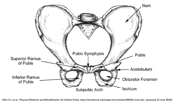
The symphysis pubic is found on the anterior side of the pelvis and is the anterior boundary of the perineum.[4][2] The Pubic bones form a cartilaginous joint in the median plane, the symphysis pubis.[2] The Joint keeps the two bones of the pelvis together and steady during activity.
In cooperation with the sacroiliac joints the symphysis forms a stable pelvic ring. This ring allows only some small mobility.[5]
The pubic symphysis is a cartilaginous joint which consists of a fibrocartilaginous interpubic disc.[2][4]The Pubic bones are connected by four ligaments. The superior pubic ligaments start on the superior part of the pubis and go as far as the pubic tubercles. The arcuate pubic ligaments form the lower border of the pubic symphysis and blends with the fibrocartilaginous disc.[2] The joint stability is mostly given by the arcuate ligament and is also the strongest one. The four ligaments together neutralise shear and tensile stresses.[4]
The disc connects the adjacent surfaces of the two pubic bones. Both of these surfaces contain a thin layer of hyaline cartilage.[2]
The junction is not flat, there are papilliform elevations with reciprocal depressions and ridges.[4]
This disc is very small in children and the hyaline cartilage is very wide, but it evolves conversely. The disc is higher, smaller and narrower in men.[2][4] Normally with women is the symphysis pubic gap 4-5 mm wide and it widens 2-3 mm during the last trimester of pregnancy. This is necessary to facilitate the delivery of the baby[4] With symphysis pubis dysfunction the joints become more relaxed and allow instability in the pelvic girdle. When the gap is equal to or more than 10 mm there’s a diastasis of the symphysis pubic.[4]
Epidemiology/Etiology
We are NOT certain what the cause is of SPD, but there are some theories:
Aslan et al. say that the etiology of SPD is unknown.[2] Pregnancy leads to an altered pelvic load, lax ligaments and weaker musculature. This leads to spino-pelvic instability, which manifest itself as symphysis pubic dysfunction.[4]
In early stages of the pregnancy the corpus luteum secretes a lot of relaxin and progesterone.[4] From the 12th week of the pregnancy this function is continued by the placenta and decidua.[4] Relaxin breaks down collagen in the pelvic joint and causes laity and softening. Progesterone has a similar effect. But relaxin has no correlation with the degree of symptoms of symphysis pubic dysfunction.[4] A Norwegian study showed that genetic susceptibility to joint dysfunction is possibly caused by an aberration of relaxin.[6] It would seem that the presence of laxity as the result of a hormonal link is undisputed.[4] However there is no full disclosure for this.
Other factors contributing to SPD include physically strenuous work during pregnancy and fatigue with poor posture and lack of exercise. Weight gain, multiparity, increased maternal age and a history of difficult deliveries, including shoulder dystocia, may also play a role.
The effective accommodation of the joints to each specific load demand through an adequately tailored joint compression, as a function of gravity, coordinated muscle and ligament forces, to produce effective joint reaction forces under changing conditions, Because of the pregnancy (hormones) ligaments and muscles become more lax and no longer have the same ability than they had before, this is why Pregnancy leads to an altered pelvic load, . In combination these lead to spino-pelvic instability, most commonly manifest as SPD.[4]
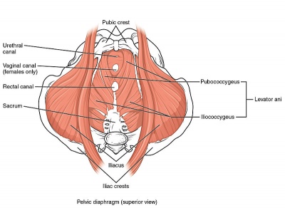
In short there the causes for this instability include hormonal (relaxin), metabolic (calcium), biomechanical (load of pregnancy or physical exercise(weak musculature)), body composition (weight), anatomical and genetic variations.[2] there are 3 layers of pelvic floor muscles, Superficial perineal layer (innervated by the pudendal nerve): Bulbocavernosus, Ischiocavernosus, Superficial transverse perineal, External anal sphincter (EAS).
Deep urogenital diaphragm layer (innervated by pudendal nerve): Compressor urethera, Uretrovaginal sphincter, Deep transverse perineal.
Pelvic diaphragm (innervated by sacral nerve roots S3-S5): Levator ani [pubococcygeus (pubovaginalis, puborectalis), iliococcygeus, coccygeus/ischiococcygeus], Piriformis, Obturator internus.
The pelvic floor muscles function as support for the organs that lie on it. The sphincters (anal and urethral sphincters) provide conscious control over the bowels and bladder, respectively, such that we are able to control the release of feces/flatus, or urine, and to prevent and delay emptying until convenient.
Upon contraction, pelvic floor muscles will lift upwards the internal organs, and tighten the sphincters openings of the vagina, anus and urethra. Relaxation of the pelvic floor muscles allows for passage of feces and urine.
Pregnancy tends to change all these muscles and so also their function.
Characteristics/Clinical Presentation
Symphysis pubic dysfunction is a condition that causes excessive movement of the pubic symphysis in the anterior or lateral direction and causes pain.
Symptoms[4]:
- Pain:
- Burning, shooting, grinding or stabbing
- Mild or prolonged
- Usually relieved by rest
- Radiating to the back, abdomen, groin, perineum and legs
- Disappears commonly after giving birth (not in every case)
- Discomfort sense onto the front of the joint
- Clicking of the lower back, hip joints and saccroillial joints when changing position
- Difficulty in movements like ab- and adduction
- Locomotor difficulty: walking, ascending or descending stairs, rising from a chair, weight bearing activities, standing on one leg, turning in bed, …
- Depression, possibly due to the discomfort
- Increased level of cases of pubic symphysis dysfunction
- 75% women developed SPD in the first trimester of pregnancy (Norwegian study) while 89% women developed SPD in the second and third trimester (UK study)
- according to Owens K. et al. injury arises in:[1][7]
- first trimester of pregnancy for 9% of women
- second trimester of pregnancy for 44% of women
- third trimester for 15% of women
- postnatal of 2% of women
- incidence of 1:36 and 1:300 in British population[7]
- occasionally, SPD may occur in labour or in the puerperium
- VAS = 7/10
Cause:[4]
- Diastasis
- Rupture
- Osteomy
- Fracture
- Misalignment of the pelvis
- Frequently associated with pregnancy and childbirth
- Increased age of getting a child
- Sports injury: caused by falling with the legs in hyper-abbduction (example: horse-riding)
- Prostatectomy
Differential Diagnosis
Leadbetter et al. found, according to their findings of a scoring system to diagnose symphysis pubic dysfunction 5 symptoms which might be significant to determine symphysis pubic dysfunction [2]
1. pubic bone pain on walking
2. standing on one leg
3. climbing stairs
4. turning over in bed
5. previous damage to lumbo-sacral spine or pelvis
The potential symptoms of the differential diagnosis of SPD need to be firmly excluded through clinical history, physical examination and appropriate investigations, to ensure the diagnosis of symphysis pubic dysfunction.
The symptoms which may lead to a diagnosis of SPD, are: nerve compression (intervertebral disc lesion), symptomatic low back pain (lumbago and sciata), pubic osteolysis, osteitis pubis, bone infection (osteomyelitis, tuberculosis, syphilis), urinary tract infection, round ligament pain, femoral vein thrombosis and obstetric complications.[2][4]
Diagnostic Procedures
As in all dysfunctions, an early diagnosis is important to minimize the possibility of a long term problem. However, not all healthcare practitioners recognise this problem.
A diagnosis is often made symptomatically e.g. after a pregnancy but imaging is the only way to confirm diastasis of the symphysis pubis.[4]Radiography, like an MRI (magnetic resonance imaging), x-ray, CT (computerised tomography) or ultrasound[1, level 1A], has been used to confirm the separation of the symphysis pubis.[4] Although it is not considered as the method of choice because of the danger of exposing the fetus to ionizing radiation. A better technique with superior spatial resolution and avoidance of ionizing radiation, is MRI.
Other techniques that may aid in diagnosis and monitoring the treatment of symphysis pelvic dysfunction, are transvaginal or transperineal ultrasonography, using high-resolution transducers.[2] Ultrasonography is a useful diagnostic aid which can measure the interpubic distance. This can be a consequence of diastasis of the pubic symphysis following childbirth.
The interpubic distance is usually measured with electronic callipers. It’s also important to know that ultrasonography provides a simple means of measuring interpubic gap, without exposure to ionizing radiation.[8]
Outcome measures
Pubic Symphysis Dysfunction has been described as a collection of signs and symptoms of discomfort and pain in the pelvic area. We still don’t know 100% what the etiology is of Pubic Symphysis Dysfunction, therefore it is not easy to create something to measure the difference between the beginning of the therapy and the end other than pain(PDI, Pain disability index) and instability in the pelvic girdle.
However there has been some research to develop a scoring system for symphysis pubis dysfunction more research is needed on this subject.
all groups showed significant improvement in function over time, as measured by the Roland-Morris Questionnaire and the Patient-Specific Functional Scale, but there was no significant difference (P .05) among groups.[9]
Examination
A physical examination is important for differentiating other possible causes, such as lumbar spine problems or prolapsed discs and to get a proper diagnosis.[2][4]
Here are some examination techniques:
Palpation[2]
- Tenderness of the symphysis pubis
- Tenderness of The sacroiliac joint
- the sacrotuberous ligament
- The tenderness of muscles including: Gluteal muscles, M. iliopsoas, M. piriformis and para-vertebralis
Provocative tests (when positive, they are helpful in diagnosing SPD) [2][10]
- Patrick’s Fabere sign
- One iliac spine is fixed by the examiner, the patient lies in a supine position, the patient places the opposite heel on the ipsilateral knee with the leg falling passively outwards. The test is positive when there is pain in either sacroiliac joint
- Active straight leg raise (ASLR)
- Pain at symphysis when standing on one leg
- Bilateral trochanteric compression
Range of motion can be limited by pain[4], Particularly during lateral rotation and during abduction.
A waddling gait[2][4], This can be the result of a tendency of the M. gluteus medius which loses its function as abductor.
A continuous pain during specific activities, can predict Pubic symphysis dysfunction.[2][4]
- Walking
- Climbing stairs
- Turning in bed
- Standing on one leg or weight
- Rising from a chair.
There are some tests for symphyseal pain in pregnancy that have a high specificity, sensitivity and inter-examiner reliability ( with a Kappa coefficient > 0.40) [4]
- Palpation with the patient in supine of the entire anterior surface of the symphysis pubis, elicits pain that stays for more than 5 seconds after the removal of the examiner’s hand. (99% specificity, 60% sensitivity and 0.89 Kappa coefficient [4]
- When you let the patient stand on one leg, they are unable to maintain their pelvis in the horizontal plane cause the opposite buttock drops (The Trendelenburg’s sign: 99% specificity, 60% sensitivity and 0.63 Kappa coefficient)[4]
- Patrick’s fabere sign, see ‘palpation’ (specificity 99%, 40% sensitivity and 0.54 Kappa coefficient)[4]
Medical management
During pregnancy:
- Paracetamol[4]
- Codeine-based preparations [4]
- Lumbar epidural morphine/bupivacaine/fentanyl usage for 24-72 hours, to break the vicious cycle of pain and muscle spasm[11]
After delivery:
- NSAIDS[4]
- Lumbar epidural morphine/bupivacaine/fentanyl usage for 24-72 hours, to break the vicious cycle of pain and muscle spasm[11]
Others:
- Hospital pain team, when intractable cases[4]
- Intra-symphyseal injection of a combination of hydrocortisone, chymotrypsin and lidocain[4]
Keep always close monitoring of effectiveness and side-effects![4]
Physical therapy management
Devices[4]
- Elbow crutches
- Pelvic support devices
- Lumbopelvic belt
- The belt must be positioned just cranial to the greater trochanters. The study advises not to use pelvic belts as a monotherapy because stability of the lumbopelvic area has to established by proper motor control and coordination.
- Prescriped pain reliefs (carefull taking NSAID while pregnant)
- Wheelchair in very severe cases
- Social services
Birth planning [4]
- Women with PSD should give birth in an upright position, with knees slightly open
- the gap may never exceeds the maximal comfort zone which is why it’s suggested that the patient wear a ribbon to both legs.[2][4]
- Practices such as placing the feet on the midwife’s hips during birth, stirrups, and interventions such as forceps should be avoided in the delivery room if possible because they can strain ligaments further.[4]
- During labour and delivery leg abduction(=separation) should be kept to a minimum[4].
Prevention[12]
- giving information
- about the condition and relationship between impairment, load demand, and actual loading capacity
- the importance of necessary rest
- to reduce fear
- to enable patients to become active in their own treatment
- Giving advice to use the body in daily life (Sit down while doing things if possible, Sleep with a pillow between the legs. Keeps legs flexed and together to get in/ out of bed)[4]
- Avoid activities that put undue strain on the pelvis (squatting, strenuous exercises, prolonged standing, lifting and carrying, stepping over things, twisting movements of the body, vacuum cleaning and stretching exercises)[4][5]
- The lumbopelvic belt combined with information is superior to exercise and information or information alone. The women are allowed to remove the belt only during sleeping.[13]
Exercises [4][5] (Therapy Exercises for the Hip)
- Aerobic exercises:
- brisk walking with a medium intensity
- defined as 64 to 76% of maximum heart rate
- for 25 minutes per day and 3 days per week
- Stretching exercises:
- hamstring, inner thigh, side waist, quadriceps and back stretch
- for 2 times per day and 3 times per week
- duration of each occasion: 10 to 20 seconds
Strengthening exercises:
- forward bending, back pressing, diagonal curling, upper body bending, leg lift crawling along with kegel exercise and pelvic tilt was given to the patients.
- for 3 to 5 times per each exercise session for both sides of the body while performing 2 exercise bouts per day and 3 days per week
- duration of each occasion: 3 to 10 seconds
- pelvic muscle floor exercises [1, level 1a] (Exercises for Lumbar Instability)
- early pregnancy: to reduce the risk to develop SPD
- deep abdominal exercises: to increases the core stability and to prevent women to develop pelvic or back pain during pregnancy
- can be progressed by starting off with a small number of repetitions and gradually increasings the seconds to hold on a contraction
- m. transversus abdominus is an important muscle, when he contracts, we can see a synergistic activation of the pelvic floor
stabilisation exercises: [4][8](Low Back Pain and Pelvic Floor Disorders) (Core stability)
- aimed at improving motor control and stability through improving force closure of the pelvis
- initially: contraction of the transveres abdominal wall muscles
- specific training of the deep local muscles like the transverse abdominal wall muscles with co-activation of the lumbar multifidus in the lumbosacral region
- training of the superficial global muscles
- Exercises to increase circulation in hip rotator muscles.[12]
- many repetitions during low force in a limited range of motion
- side lying position with a pillow between the legs or sitting without foot support
other therapies[4]:
- accupunture
- TENS
- Ice
- External heat
- Massage
Their efficacy isn’t yet proven.[2][4] The effects of a treatment of a chiropractor or the effects of talking to “hands on experts” can be useful.[4]
Clinical bottom line
Pubic Symphysis Dysfunction has been described as a collection of signs and symptoms of discomfort and pain in the pelvic area, including pelvic pain radiating to the upper thighs and perineum. It can be examined by palpation, provocation tests, a wadding gait, also a continuous pain during activities can predict pubic symphysis dysfunctio. Also MRI can diagnose this condition. In latest literature we see that Pubic symphysis dysfunction is treated in a lot of ways. First there is birth planning, in this aspect several criteria could help avoid this condition. Giving information about the condition itself may be used so that the patiënt knows what can be done and what is not. Exercise is also an important aspect, where aerobic, strengthening, pelvic muscle floor and stabilisation exercises are combined. Other therapies like accupunture, TENS, ice, external heat and massage are also done but their effectivity are not yet been proven. During and after pregnancy a symptomatic treatment can be given, this is to limit the pain and is always with physical therapy.
Recent Related Research (from ● Haakstad, Lene AH, and Kari Bø. “Effect of a regular exercise programme on pelvic girdle and low back pain in previously inactive pregnant women: a randomized controlled trial.” Journal of rehabilitation medicine 47.3 (2015): 229-234.
References
- ↑ 1.01.11.2 Welsh A., Antenatal care routine care for the healthy pregnant woman, National Collaborating Centre for Women’s and Children’s Health, 2008, 2nd edition, London, 113
- ↑ 2.002.012.022.032.042.052.062.072.082.092.102.112.122.132.142.152.162.172.182.19 Aslan A. and Fynes M., Symphyseal pelvic dysfunction, Current Opinion Obstetrics and Gynecology, 2007 19:133–139.
- ↑ Leadbetter, R. E., D. Mawer, and S. W. Lindow. “Symphysis pubis dysfunction: a review of the literature.” The Journal of Maternal-Fetal & Neonatal Medicine 16.6 (2004): 349-354.
- ↑ 4.004.014.024.034.044.054.064.074.084.094.104.114.124.134.144.154.164.174.184.194.204.214.224.234.244.254.264.274.284.294.304.314.324.334.344.354.364.374.384.394.404.414.424.434.444.454.464.47 Jain, Smita, et al. “Symphysis pubis dysfunction: a practical approach to management.” The Obstetrician & Gynaecologist 8.3 (2006): 153-158.
- ↑ 5.05.15.25.3 Kordi R. Et. Al. Comparison between the effect of lumbopelvic belt and home based pelvic stabilizing exercise on pregnant women with pelvic girdle pain; a randomized controlled trial. J Back Musculoskelet Rehabil. 2013;26(2):133-9
- ↑ MacLennan AH, MacLennan SC., Symptom-giving pelvic girdle relaxation of pregnancy, postnatal pelvic joint syndrome and development dysplasia of the hip., Acta Obstet Gynecol Scand, 1997;76:760–64.
- ↑ 7.07.17.2 Owens K, Pearson A, Mason G. Symphysis pubis dysfunction – a cause of significant obstetric morbidity. Eur J Obstet Gynecol Reprod Biol 2002;105:143–46.
- ↑ 8.08.1 M.W. Scriven MS FRCS, Dai Anthony Jones MChlr FRCS, Liam McKnight FRCR, “The importance of pubic pain following childbirth: a clinical and ultrasonographic study of diastasis of the pubic symphysis, J R Soc Med 1995;88:28-30.
- ↑ Depledge J. et al., Management of Symphysis Pubis Dysfunction During Pregnancy Using Exercise and Pelvic Support Belts, Physical Therapy,2005, 1290-1300
- ↑ Albert, H. Et Al. “Evaluation of clinical tests used in classification procedures in pregnancy-related pelvic joint pain.” European Spine Journal 9.2 (2000): 161-166)
- ↑ 11.011.1 Scicluna JK, Alderson JD, Webster VJ, Whiting P. Epidural analgesia for acute symphysis pubis dysfunction in the second trimester. Int J Obstet Anesth 2004;13:50–2.
- ↑ 12.012.1 Elden H, Ladfors L, Olsen MF, Ostgaard HC, Hagberg H., Effects of acupuncture and stabilising exercises as adjunct to standard treatment in pregnant women with pelvic girdle pain: randomised single blind controlled trial, BMJ ,2005;330:761
- ↑ Stuge B, Msc, et al., The Efficacy of a Treatment Program Focusing on Specific Stabilizing Exercises for Pelvic Girdle Pain After Pregnancy, SPINE Volume 29, Number 10, pp E197–E203 2004


