Description
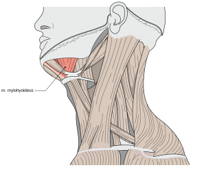
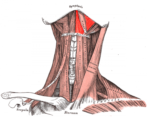
The Mylohyoideus (Mylohyoid muscle), flat and triangular, is situated immediately above the anterior belly of the Digastricus, and forms, with its fellow of the opposite side, a muscular floor for the cavity of the mouth. It arises from the whole length of the mylohyoid line of the mandible, extending from the symphysis in front to the last molar tooth behind. The posterior fibers pass medialward and slightly downward, to be inserted into the body of the hyoid bone. The middle and anterior fibers are inserted into a median fibrous raphe extending from the symphysis menti to the hyoid bone, where they join at an angle with the fibers of the opposite muscle. This median raphe is sometimes wanting; the fibers of the two muscles are then continuous. [1]
Variations
It may be united to or replaced by the anterior belly of the Digastricus; accessory slips to other hyoid muscles are frequent. [1]
Origin
Mylohyoid line on internal aspect of mandible
Insertion
Anterior three quarters: midline raphe. posterior quarter: superior border of body of hyoid bone
Nerve
Mylohyoid nerve
A motor branch of the inferior alveolar nerve, a branch of the mandibular division of the trigeminal nerve (V).
Artery
Sublingual Artery
Function
Elevates hyoid bone, supports and raises floor of mouth. Aids in mastication and swallowing
Clinical relevance
Conditions that can afflict the mylohyoid muscle include:
- Overuse injuries,
- Tears,
- Strains,
- Myopathy,
- Atrophy,
- Infectious myositis,
- Neuromuscular diseases,
- Lacerations
- Contusions
It may be responsable of commons syntomps:
Mylohyoid boutonniere
A normal focal discontinuity in the mylohyoid muscle, which may permit the sublingual salivary gland, fat or vessels – or a combination of each – to protrude out from the sublingual space into the submandibular space. [4]
Salivary gland disease
Due to the position of the submandibular gland a referred pain at the mylohyoid muscle could be present.
The submandibular gland consists of two lobes—superficial and deep—which are folded over each other posteriorly around the posterior free border of the mylohyoid muscle. [5]
Assessment
Palpation
On intra- and extraoral palpation, find a sheet of muscle attached to the whole length of the mylohyoid line of the mandible and extending to the body of the hyoid bone.[6] Use the index finger to slide all over different sheets. Continuous compression should be applicate less than 20sec.
Treatment
Resources
See also
Feeding and the Swallow Mechanism
References
- ↑ 1.01.1 Gray, Henry. Anatomy of the Human Body. Philadelphia: Lea e Febiger, 1918; Bartleby.com, 2000.
- ↑ Kenhub. Mylohyoid muscle – Origin, Insertion, Innervation, Function Definition – Human Anatomy Available from: ↑ prof.Dr Ahmed M.Kamal. 51 Mylohyoid muscle. Available from: ↑ Radiopedia. Mylohyoid Boutonierre. ↑ J.D. Langdon, Oral and Maxillofacial Surgery (Second Edition). In: Salivary gland disease. Elsevier, 2007. Pages 199–214
- ↑ Torsten Liem, Cranial Osteopathy (Second Edition). In: Chapter 12 – The orofacial structures, pterygopalatine ganglion and pharynx. Elsevier, 2004. Pages 437-484


