Phrenic Nerve
The Phrenic nerve is a nerve of the thoracic region. It is important for breathing, as it passes motor information to the diaphragm and receives sensory information from it. There are two phrenic nerves, a left and a right one.[1]
Function
Both of these nerves supply motor fibers to the diaphragm and sensory fibers to the fibrous pericardium, mediastinal pleura, and diaphragmatic peritoneum.[1]
Origin
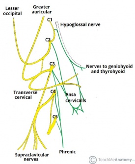
The phrenic nerve originates mainly from the 4th cervical nerve, but also receives contributions from the 3rd and 5th cervical nerves (C3-C5) in humans.[2] Thus, the phrenic nerve receives innervation from parts of both the cervical plexus and the brachial plexus of nerves.
Course
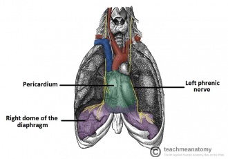
The right Phrenic nerve descends in the thorax along the right side of the right brachiocephalic vein and the superior vena cava. It passes in front of the root of the right lung and runs along the right side of the pericardium, which separates the nerve from the right atrium. It then descends on the right side of the inferior vena cava to the diaphragm.Its terminal branches pass through the caval opening in the diaphragm to supply the central part of the peritoneum on its under aspect. (Image 2)
The left Phrenic nerve descends in the thorax along the left side of the left subclavian artery. It crosses the left side of the aortic arch and here crosses the left side of the left Vagus nerve. It passes in front of the root of the left lung and then descends over the left surface of the pericardium which separates the nerve from the left ventricle. On reaching the diaphragm, the terminal branches pierce the muscle and supply the central part of the peritoneum on its under aspect.(Image 2)[1]
Motor Supply
The phrenic nerves possess efferent and afferent fibres. The efferent fibres are the sole nerve supply to the muscle of the diaphragm. The phrenic nerve supplies the ipsilateral diaphragm.[3]
Sensory supply
The afferent fibres carry sensation to the central nervous system from the peritoneum covering the central region of the under-surface of the diaphragm, the pleura covering the central region of the upper surface of the diaphragm, and the pericardium and mediastinal parietal pleura.[3]
Relations in the Neck
Anteriorly : The preverterbral layer of deep fascia, the internal jugular vein, the superficial cervical and suprascapular arteries and, on the left, thoracic duct; the beginning of the brachiocephalic vein.
Posteriorly : The scalenus anterior, the subclavian artery, and the cervical dome of pleura.
Paralysis of the Diaphragm
The phrenic nerve may be paralysed because of pressure from malignant tumours in the mediastinum. Surgical crushing or sectioning of the phrenic nerve in the neck, producing paralysis of the diaphragm on one side, was once used as a part of the treatment of lung tuberculosis, especially of the lower lobes. The immobile dome of the diaphragm rests the lung.[4]
References
- ↑ 1.01.11.2 Prakash; Prabhu, L. V.; Madhyastha, S; Singh, G (2007). ↑ Moore, Keith L. (1999). Clinically oriented anatomy. Philadelphia: Lippincott Williams & Wilkins. ISBN 978-0-683-06141-3
- ↑ 3.03.1 Loukas M, Kinsella Jr CR, Louis Jr RG, Gandhi S, Curry B. Surgical anatomy of the accessory phrenic nerve. The Annals of thoracic surgery. 2006 Nov 1;82(5):1870-5.
- ↑ Yu-Dong, G., Min-Ming, W., Yi-Lu, Z., Jia-Ao, Z., Gao-Meng, Z., De-Song, C., Ji-Geng, Y. and Xiao-Ming, C. (1989), Phrenic nerve transfer for brachial plexus motor neurotization. Microsurgery, 10: 287–289. doi: 10.1002/micr.1920100407


