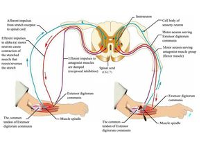
Spinal Reflex/The Reflex Arc
A reflex is an involuntary and nearly instantaneous movement in response to a stimulus. The reflex is an automatic response to a stimulus that does not receive or need conscious thought as it occurs through a reflex arc. Reflex arcs act on an impulse before that impulse reaches the brain.[1]
Relex arcs can be
- Monosynaptic ie contain only two neurons, a sensory and a motor neuron. Examples of monosynaptic reflex arcs in humans include the patellar reflex and the Achilles reflex.
- Polysynaptic ie multiple interneurons (also called relay neurons) that interface between the sensory and motor neurons in the reflex pathway.[2]


The video below illustrates the reflex arc
Reflex Testing
Deep Tendon (muscle stretch) Reflexes
Evaluates afferent nerves, synaptic connections within the spinal cord, motor nerves, and descending motor pathways. Lower motor neuron lesions (eg affecting the anterior horn cell, spinal root or peripheral nerve) depress reflexes: upper motor neuron lesions increase the reflexes.
Reflexes tested include the following:
- Biceps (innervated by C5 and C6)
- Radial brachialis (by C6)
- Triceps (by C7)
- Distal finger flexors (by C8)
- Quadriceps knee jerk (by L4)
- Ankle jerk (by S1)
- Jaw jerk (by the 5th cranial nerve)
Technique for testing reflexes
- The muscle group to be tested must be in a neutral position (i.e. neither stretched nor contracted).
- The tendon attached to the muscle(s) which is/are to be tested must be clearly identified. Place the extremity in a positioned that allows the tendon to be easily struck with the reflex hammer.
- To easily locate the tendon, ask the patient to contract the muscle to which it is attached. When the muscle shortens, you should be able to both see and feel the cord like tendon, confirming its precise location.
- Strike the tendon with a single, brisk, stroke. You should not elicit pain.
This grading system is rather subjective.
- 0 No evidence of contraction
- 1+ Decreased, but still present (hypo-reflexic). Hyporeflexia is generally associated with a lower motor neuron deficit (at the alpha motor neurons from spinal cord to muscle) eg Guillain–Barré syndrome
- 2+ Normal
- 3+ Super-normal (hyper-reflexic) Hyperreflexia is often attributed to upper motor neuron lesions eg Multiple sclerosis
- 4+ Clonus: Repetitive shortening of the muscle after a single stimulation[4]
Note any asymmetric increase or depression. Jendrassik manoeuvre can be used to augment hypoactive reflexes ie the patient locks the hands together and pulls vigorously apart as a tendon in the lower extremity is tapped or can push the knees together against each other, while the upper limb tendon is tested.
The video below illustrates the testing of the deep tendon reflexes
Pathologic reflexes
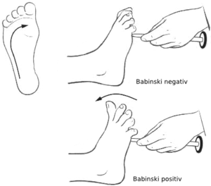
Pathologic reflexes (eg, Babinski, rooting, grasp) are reversions to primitive responses and indicate loss of cortical inhibition.
Other reflexes
Clonus (rhythmic, rapid alternation of muscle contraction and relaxation caused by sudden, passive tendon stretching) testing is done by rapid dorsiflexion of the foot at the ankle. Sustained clonus indicates an upper motor neuron disorder.[6]
References
- ↑ Wikipedia. ↑ Lumen. ↑ Dr Matt and Dr Mikes Medical youtube. Spinal reflex Available from: ↑ University of California. ↑ Justin Vaida Deep tendon reflexes. Available from: ↑ MSD Manual. function gtElInit() { var lib = new google.translate.TranslateService(); lib.setCheckVisibility(false); lib.translatePage('en', 'pt', function (progress, done, error) { if (progress == 100 || done || error) { document.getElementById("gt-dt-spinner").style.display = "none"; } }); }
Ola!
Como podemos ajudar?


