Introduction
All health care professionals are aware of this term “Babinski Sign”. It is an essential part of any Neuro-Assessment. As any other procedure, this sign is also topic of debate for a long time. However its use in clinical routine remains unabated.
History
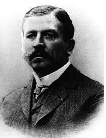
It was on February 22, 1896, that Joseph Francois Felix Babinski published his first report on ‘reflexe cutane plantaire’ [cutaneous plantar reflex] which became the sign that bears his name: ‘the Babinski sign’.[1] He referred to the sign as “phénomène des orteils” (toes phenomenon) but is now usually referred to eponymously as the “Babinski sign” or descriptively as the extensor plantar response.
Babinski Sign
This eponym refers to the dorsiflexion of the great toe with or without fanning of the other toes and withdrawal of the leg, on plantar stimulation in patients with pyramidal tract dysfunction.
Neuro-Physiology
The neurophysiology of this reflex has not been completely elucidated.
- Each area of the skin of the body appears to have a specific reflex response to noxious stimuli. The purpose of the reflex is to cause the withdrawal of the area of the skin from the stimulus.
- This reflex is mediated by the spinal cord, but influenced by higher centers. The area of skin from which the reflex can be obtained is known as the receptive field of the reflex. [2]
- The abnormal plantar reflex, or Babinski reflex, is the elicitation of toe extension from the “wrong” receptive field, that is, the sole of the foot. Thus a noxious stimulus to the sole of the foot produces extension of the great toe instead of the normal flexion response.
- The essential phenomenon appears to be recruitment of the extensor hallucus longus, with consequent overpowering of the toe flexors .[3]The movements of the other joints remain the same.
- The corticospinal tract influences the segmental reflex in the spinal cord. When the corticospinal tract is not functioning properly, the result is that the receptive field of the normal toe extensor reflex enlarges at the expense of the receptive field for toe flexion. Toe extension is consequently elicited from what is normally the receptive field for toe flexion.
- The maintenance of territorial integrity of the receptive fields is apparently one way in which the cortex exerts its influence under normal conditions.
Non- Neurological Causes of Extensor Plantar Response[1]
- In children upto the age of 1 year
- Deep sleep
- Coma
- General anaesthesia
- Electroconvulsive therapy
- Post-ictal stage of epilepsy
- Apnoeic phase of Cheyne Stockes breathing
- Narcosis
- Alcohol intoxication
- Hypoglycemia
- Hypnosis
- Physical exhaustion and marathon walking
- Drugs-scopalamine, barbiturates
Technique
Instruction to the patient:
Place the patient in a supine position and tell him or her that you are going to scratch the foot.
Handling:
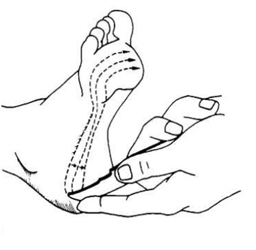
Fixate the foot by grasping the ankle or medial surface with the examiner’s hand that will be closest to the midline of the patient: examiner’s left hand when the patient’s left foot is being tested, and vice versa with the right foot.
Procedure:
The first line to be stroked begins a few centimeters distal to the heel and is situated at the junction of the dorsal and plantar surfaces of the foot. The line extends to a point just behind the toes and then turns medially across the transverse arch of the foot. Stroke slowly, taking 5 or 6 seconds to complete the motion. Do not dig into the sole, but stroke.
Clinical Implication
- The plantar reflex is a nociceptive segmental spinal reflex that serves the purpose of protecting the sole of the foot.
- The clinical significance lies in the fact that the abnormal response reliably indicates metabolic or structural abnormality in the corticospinal system upstream from the segmental reflex.
- Thus the extensor reflex has been observed in structural lesions such as hemorrhage, brain and spinal cord tumors, and multiple sclerosis, and in abnormal metabolic states such as hypoglycemia, hypoxia, and anesthesia.[2]
Judging the response
The normal and pathological responses to plantar stimulation are succinctly described by Babinski in his original communication [4]
“On the healthy side pricking of the sole provokes… flexion of the thigh on the pelvis, of the leg on the thigh, of the foot on the leg and of the toes upon the metartasus. On the paralysed side a similar excitation also results in flexion of the thigh on the pelvis, of the leg on the thigh, of the foot on the leg, but the toes, instead of flexing, execute a movement of extension upon the metatarsus.”
These rules have been shown to improve accuracy of the sign when compared against clinical and electromyographic recordings[5]
- Upward movement of the toe is pathological only if caused by contraction of extensor hallicus longus muscle
- Contraction of extensor hallicus longus muscle is pathological only if it occurs synchronously with reflex activity in other flexor muscles
- A true up-going toe sign is reproducible, unlike voluntary withdrawal.
Potential causes of an incorrectly interpreted positive Babinski sign[6]
- Contraction of tibialis anterior: This causes the toes to go up passively due to ankle movement without contraction of EHL
- Very active flexion synergy: Following a brisk normal flexion synergy reflex including usual flexion of the toes, the observer sees the big toe return to its neutral position by going up, but this is caused by relaxation of the toe flexors rather than EHL contraction
- Voluntary toe wriggling: These movements are jerky, inconsistent and not synchronous to flexion synergy in the rest of the leg
- Relative movement: The smaller toes go down, whilst the big toe remains immobile creating the illusion of an up-going toe
- ‘Isolated fanning of the toes’: Although suggested by some to be an important feature of the Babinski sign – in fact may be seen in healthy subjects and may not be seen in Babinski sign so is of limited use
Red Alert
An extensor response may be present when there is no damage to the pyramidal tract.
A possible explanation being the excitation of the distal motor neurons and inhibition of the impulses via flexor reflex afferent nerve fibres can be dissociated because they are mediated by different neurons, however closely linked. [2]
On the contrary, cases with proven damage to the pyramidal system have had normal plantar response.
The corticospinal fibres not only originate in different parts of the cortex, but also have different terminations. Babinski sign can be expected only when ‘leg fibres’ of the pyramidal tract are involved.
References
- ↑ 1.01.1 S.P. Kumar, D. Ramasubramanian, The Babinski Sign – A Reappraisal, REVIEW ARTICLE, Neurol India, 2000; 48 : 314-318
- ↑ 2.02.12.2 H. KENNETH WALKER, The Plantar Reflex, In:The Neurological System, Pg-369-370.
- ↑ Landau W. Clinical definition of the extensor plantar reflex (letter) .N Engl J Med 1971 ;285 :1149-50 .
- ↑ Babinski J (1896) Sur le reflexe cutane plantaire dans certaines affections du systeme nerveux central. Comptes rendus des Seances et Memoires de la Societe de Biologie, 207-208
- ↑ van Gijn J (1976) Equivocal plantar responses: a clinical and electromyographic study. J. Neurol. Neurosurg. Psychiatr 39, 275-282
- ↑ Jasper M Morrow, Mary M Reilly , The Babinski sign, MRC Centre for Neuromuscular Diseases, Department of Molecular Neuroscience, UCL Institute of Neurology,


