Definition/Description
Posterior interosseous nerve syndrome is a neuropathic compression of the posterior interosseous nerve where it passes through the radial tunnel.[1] This may result in paresis or paralysis of the digital and thumb extensor muscles, resulting in an inability to extend the thumb and fingers at their metacarpophalangeal joints. The only movement patients may be able to do is the dorsoradial direction.[2]
Clinically relevant anatomy
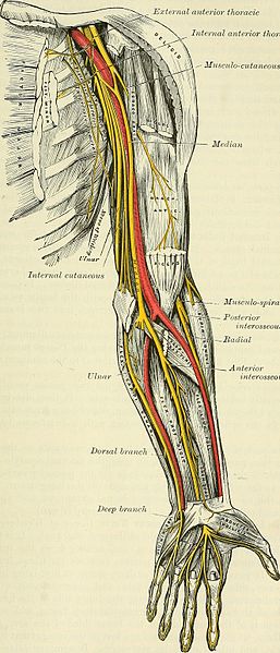
The posterior interosseous nerve is located close to shaft of the humerus and the elbow. This nerve is the deep motor branch of the radial nerve. Proximal to the supinator arch, the radial nerve is divided into a superficial branch and posterior interosseous branch.The radial nerve supplies the majority of the forearm and hand extensors. Damage to this branch of the radial nerve results in posterior interosseous nerve syndrome.
The radial tunnel is a space that extends 5cm from the radial head to the distal margin of the supinator. This tunnel is attached laterally to the brachioradialis, extensor carpi radialis longus and extensor carpi radialis brevis and medially to the biceps tendon and brachialis. The floor is formed by the deep head of the supinator and the capsule of the radiocapitellar joint, while the roof is formed by the superficial head of the supinator and the radial recurrent vessels.[3]
At the level of the lateral epicondyle, between the brachioradialis and brachialis muscles, the radial nerve, which has its origin in the brachial plexus, divides into its 2 terminal branches: the superficial radial nerve and the posterior interosseous nerve.[4] The superficial radial nerve ends proximal to the radial tunnel. The posterior interosseous nerve is much longer and enters the radial tunnel underneath a musculotendinous arch, the arcade of Frohse. The arcade of Frohse, which is the most common point of compression, is a connection between the deep and superficial heads of the supinator and is fibrotendinous in 30% of the population. The posterior interosseous nerve continues in the radial tunnel through the supinator, as it goes from the anterior to the posterior surface of the forearm.
The posterior interosseous nerve is a motor nerve and sequentially innervates supinator, extensor carpi radialis brevis, extensor digitorum communis, extensor digiti minimi, extensor carpi ulnaris, abductor pollicis, extensor pollicis brevis, extensor pollicis longus, and extensor indicis.[3][5]
Epidemiology/Etiology
Epidemiology
Posterior interosseous nerve syndrome is more common in males, manual laborours and bodybuilders, with an incidence of 3 per 100 000.[6] With a humeral shaft fracture, there is a 12% chance of associated with radial nerve paralysis.[7]
Etiology
Posterior interosseous nerve syndrome can be caused by a traumatic injury, tumors, inflammation and an anatomic injury. With repeated pronation and supination a dynamic compression of the nerve in the proximal part of the forearm can be created.[8]
Posterior interosseous nerve syndrome usually develops spontaneously[1] and is caused by compression injuries to the upper extremity, mostly in the arcade of Frohse[9]. It is the area where the nerve enters the supinator muscle[10] and is the most common place for a compression of the nerve. However, it can also occur following trauma, such as a blow to the proximal dorsal region of the forearm. Impingement of the radial nerve results in posterior interosseous nerve syndrome.[7] Compression of the posterior interosseous nerve is associated with repetitive activities that involve wrist supination and pronation, with a component of wrist extension.[11]
Posterior interosseous nerve syndrome can be iatrogenic following reduction of radial fracture, transposition of the ulnar nerve or release of the extensor origin for lateral epicondylitis.[1] The causes of posterior interosseous nerve syndrome include intrinsic nerve abnormalities and extrinsic compression.[2]
Characteristics/Clinical presentation
Most nerve entrapments occurs due to an osseoligamentous tunnel narrowing. In the case of a posterior interosseous nerve entrapment, the compression occurs within the musculo-tendinous radial tunnel. In 69.4%, the nerve is compressed by the fibrous arcade of Frohse.[12]
There is a very slow development of the symptoms. The duration of symptoms averaged 2-3 years before a definitive diagnosis could be made.[8] Symptoms of nerve entrapment syndromes are generally involving pain, sensory and motor changes, sensations of popping, paresthesias, and paresis.
Posterior interosseous nerve syndrome is characterized by motor deficits in the distribution of the posterior interosseous nerve.[12] While the posterior interosseous nerve does have afferent fibres that transmit pain signals from the wrist, it does not carry any cutaneous sensory information that can help distinguish a posterior interosseous nerve palsy from Differential diagnosis
Posterior interosseous nerve syndrome is one of the pathologies that can cause lateral elbow pain. The other pathologies that are associated with lateral elbow pain are:
Diagnostic procedures
Careful clinical and electrophysiological examination is important and essential for a reliable diagnosis.[10]
Physical examination
- History
- Functional limitations or deficits
- Palpation: Abnormal tenderness is expected over the arcade of Frohse and eventually over the lateral epicondyle
- Neural tension test
- Muscle testing (with resistance):[3][15] There a partial or complete paralysis of the wrist extensors:
- The patient is unable to extend the thumb and other fingers of the affected side at the metacarpophalangeal joints
- Wrist extension is possible, but only with a dorso-radial direction, due to the weakened extensor carpi ulnaris
- Resisted supination and pronation of the forearm can produce pain, as well as resisted extension of the middle finger
- The brachioradialis, extensor carpi radialis longus and extensor capri radialis brevis are innervated by more proximal branches of the radial nerve, so may be spared
Special investigations
The following special investigations are used to assist in making the diagnosis.[1] It further aids to establish the topography of the lesion and the severity of the muscular denervation.[10]
- Electromyography: Identify level of compression
- Nerve conduction velocity
- MRI: Not commonly used:
- To determine specific area of compression
- Assist in surgical planning
Outcome measures
Medical management
There are several medical ways to treat the posterior interosseous nerve syndrome.
Conservative management
- Reduction of local inflammation and swelling around the nerve:[16]
- Wrist and/or elbow splints
- The arm can be put in an above-elbow cast for ten days with the elbow flexed at 90°, the forearm supinated and the wrist in neutral position[17]
- NSAID’s
- Activity modification to reduce local inflammation and swelling around the nerve
- Wrist and/or elbow splints
- Corticosteroid injections[17]
- Therapeutic ultrasound[17]
- Physiotherapy[17]
- Reduction of synovitis:[18]
- Heat
- Rest
- Mild range of motion
Surgery
- Indication:
- No improvement with conservative management
- Pain present after 12 weeks
- Aim: To obtain full recovery
- Surgery: Depends on how and where impingement is present[18][19]
- Arcade of Frohse release
- Resection of lesions
- Posterior interosseous nerve release
Physiotherapy management
Conservative management
3-6 months of physiotherapy with regular re-assessment of signs and symptoms is recommended. If there is no response to therapy, evidence of denervation, or persistent paralysis, surgical decompression should be considered.[12]
Physiotherapy should include a multimodal approach. The following can be considered based on the patient presentation:
- Cryotherapy: Increase extensibility and reduce tone of local muscles
- Ultrasound
- TENS
- Deep tissue massage and stretching exercises: Improve extensibility of the muscles who surround the brachial plexus and radial nerve
- Dry needling: Increase extensibility and reduce tone of local muscles
- Neural mobilizations:[20]
- Reduce mechanical extra and intra-neural adhesion
- Assist the neuromodulation of symptoms
- Manual therapy[7]: Regain elbow mobility
- Strengthening[12] and range of motion exercises
- Stretching exercises:
- Focus on supinator
- Passive wrist extensions stretches:
- Place hand on table and move upper body over wrist
- Prayer stretch
Post-surgical rehabilitation
- Commence active range of motion from day 3-5
- Incorporate stretching of extensors
- Commence strengthening from week 3-4
Patients can return to light duty work between week 2 and 3 post-operatively, while return to baseline function can take between 6 and 12 weeks.
Clinical bottom line
The posterior interosseous nerve is the deep branch stemming off the radial nerve. Compression can be caused by trauma, repetitive strain and inflammation. This is then known as posterior interosseous nerve syndrome, which may result in paresis or paralysis of the digital and thumb extensor muscles, resulting in an inability to extend the thumb and fingers at their metacarpophalangeal joints. Conservative management includes splinting, NSAID’s and physiotherapy, and symptoms normally resolve within 3-6 months. Failed conservative management is an indication for surgery, where nerve releases are the most common surgical intervention. Physiotherapy also plays a big part in the post-operative management, and rehabilitation generally lasts between 6 and 12 weeks.
References
- ↑ 1.01.11.21.31.4 Vrieling C, Robinson PH, Geertzen JH. ↑ 2.02.1 Chien AJ, Jamadar DA, Jacobson JA, Hayes CW, Louis DS. ↑ 3.03.13.2 Cha J, York B, Tawfik J. Posterior interosseous nerve compression. Eplasty 2014;14.
- ↑ 4.04.14.24.3 Bevelaqua AC, Hayter CL, Feinberg JH, Rodeo SA. ↑ 5.05.15.2 Ekstrom RA, Holden K. ↑ Ortho Bullets. PIN Compression Syndrome. Available from: ↑ 7.07.17.2 Quignon R, Marteau E, Penaud A, Corcia P, Laulan J. ↑ 8.08.1 Molina AP, Bour C, Oberlin C, Nzeusseu A, Vanwijck R. ↑ Andreisek G, Crook DW, Burg D, Marincek B, Weishaupt D. ↑ 10.010.110.2 Huisstede BM, Miedema HS, Van Opstal T, De Ronde MT, Kuiper JI, Verhaar JA, Koes BW. ↑ Rosenbaum R. Disputed radial tunnel syndrome. Muscle & Nerve: Official Journal of the American Association of Electrodiagnostic Medicine 1999;22(7):960-7.
- ↑ 12.012.112.212.3 Saratsiotis J, Myriokefalitakis E. ↑ 13.013.1 Millender LH, Nalebuff EA, Holdsworth DE. ↑ 14.014.114.2 Kaswan S, Deigni O, Tadisina KK, Totten M, Kraemer BA. Radial tunnel syndrome complicated by lateral epicondylitis in a middle-aged female. Eplasty 2014;14.
- ↑ Singh VA, Michael RE, Dinh DB, Bloom S, Cooper M. ↑ Mansuripur PK, Deren ME, Kamal RO. ↑ 17.017.117.217.3 Maffulli N, Maffulli F. ↑ 18.018.1 Chang LW, Gowans JD, Granger CV, Millender LH. ↑ Hashizume H, Nishida K, Nanba Y, Shigeyama Y, Inoue H, Morito Y. ↑ 20.020.1 Molloy J, Neville V, Woods I, Speedy D. ↑ The Student Physical Therapist. Posterior interosseous nerve syndrome. Available from: function gtElInit() { var lib = new google.translate.TranslateService(); lib.setCheckVisibility(false); lib.translatePage('en', 'pt', function (progress, done, error) { if (progress == 100 || done || error) { document.getElementById("gt-dt-spinner").style.display = "none"; } }); }

