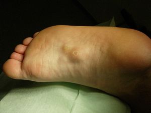
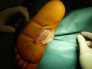
Definition
The Ledderhose disease, also known as a plantar fibromatosis or Morbus Ledderhose, is a small slow-growing lesion of the superficial fibromatoses of the plantar aponeurosis.[1] It can be described as a benign fibroblastic proliferative disorder in which fibrous nodules may develop in the plantar aponeurosis, more specifically on the medial plantar side of the foot arch and on the forefoot region.[2]
Clinically revelant anatomy
The [3]
See the page for foot anatomy for a more detailed explanation.
Epidemiology/Etiology
Ledderhose’s disease, is named after a German surgeon, Dr. Georg Ledderhose. He first described the condition in 1894 as an uncommon hyperproliferative plantar aponeurosis.[4][5]
It occurs less often than palmar disease, with a prevalence of 0.23% and usually more frequently in middle aged male individuals (30 – 50 years). Studies have shown that even though male predisposition is bigger than in females, among children there seem to be a female predilection.[1]
Due to the lack of information about the formation of this condition, the etiology is still controversial.[6] It seems to have a multifactorial etiology, including congenital and traumatic causes as well as prolonged immobilization followed by trauma.[1][7] Patients with the diabetes mellitus, epilepsy, alcoholics with liver disease, and keloids have a higher risk to develop the disease of Ledderhose and/or a Peyronie’s disease.[1][2]
Characteristics/Clinical presentation
There will be a visible bulge at the plantar area of the foot as well as a reduced capacity of bending the foot.[8][9][10][11] Not all patients have symptomatic complains. Complains such as pain can occur after standing or walking for a long time, or when those nodules happen to grow and stiffen the affected structures of the foot (due to a lack of space) such as neurovascular bundles, muscles or tendons.[6] Contractures of the foot are not expected and patients do frequently have normal radiographs.[6]
Phases
- Proliferative phase: Nodular fibroblastic proliferation
- Active phase: Collagen synthesis and deposition
- Mature phase: Reduced fibroblastic activity and collagen maturation
Differential diagnosis
Lesions may also appear at different locations of the body, such as:[3][5][8]
Diagnostic procedures
Lesions, extension, characteristics, structures involved and local recurrance can be identified on the following scans:[1][3]
- Ultrasound
- MRI: Well-defined nodule tcontinuous with the plantar fascia; Low signal intensity on T1-weighted sequences; Low to intermediate signal intensity on T2-weighted sequences
- CT scan: Used for tissue comparison; identify tissue mass (non-specific) in characteristic area; attenuation equal or higher than in skeletal muscles
Medical Management
Even though a recovery with a non-invasive treatment is possible, the severity of the lesion may demand a different approach. The most functional surgery seems to consist of a large or partial fasciectomy (in order to release the tension) with or without grafts.[10][12] Surgical removal of the nodules can also be done.[10][12]
Radiotherapy is also an option,[2]but it will mostly be used post-operatively in order to reduce the recurrence of the nodules.[5]
Physiotherapy management
The treatment of the Ledderhose disease is the same as the treatment used to rehabilitate a person that suffers from a foot.[12]
Mild cases
The treatment of a mild case of Ledderhose disease and treatment before a clinical intervention consist of:[6][12]
- Massage using cortisone cream
- Gentle passive stretching of the retracted structures
- Isometric exercises of the toe extensors
- Electrotherapy
- Elastic extension splints (here occupational therapists can also assist) – not often used
- Symptom relief by footwear modifications, pads and orthotics
- Shockwave therapy: Reducing the pain and soften the nodules
Serious cases
In serious cases, Ledderhose disease will first have to be clinically cleared, before initiating physiotherapy treatment.
Phase 1 (day 1 – 3)
- Foot should be bandaged or plastered in a functional position
- In a graft was used: Immobilize 10 days with a compressive splint-dressing
- Mobilization will only start once there is a ¾ engraftment.
- Mainly this phase will consist of:
Phase 2 (wound healing phase, day 8 – 15)
- Prolong mobilization of the free articulations
- Circulatory drills
- Scar massage
- Lymphatic drainage
- Pneumatic/air pressure therapy
- Recovery of the articulations and muscles of the toes:
- Slow passive mobilization outside of pain
- Active mobilization
- Elastic constraint switch gea
- Posture extension),
- Laser and ultrasound if challenges with would healing occurs
- Passive stretching of toe extensor foot in plantar flexion
[12][13] Phase 3
- Circulatory drills
- Scar massage
- Heat (hot water or paraffin bath) while doing active movements
- Total recovery of the articular amplitudes (by using analytic and global active-passive exercises and postural extension if needed with a dynamic brace)
- Recovery of the muscle power (manually and later on with growing mechano-therapy appliances)
- Functional training
- Home exercise program:[13][14]
- Towel curls
- Marbles pick up:
- Place some marbles on the ground next to a dish or cup and ask the patient to grip the marbles with the injured foot, one by one, in order to drop them in the cub or dish. Once this is exercise has been done, the same exercise could be accomplish with the other (not injured) side.
- Golf ball roll:
- Massage the ball of your feet by rolling the golf ball under your foot during 2 minutes.
References
- ↑ 1.01.11.21.31.4 Murphey MD, Ruble CM, Tyszko SM, Zbojniewicz AM, Potter BK, Miettinen M. ↑ 2.02.12.2 Knobloch K, Vogt PM. ↑ 3.03.13.23.3 Griffith JF, Wong TY, Wong SM, Wong MW, Metreweli C. ↑ Right Diagnosis from Healthgrades. Ledderhose disease. Available from ↑ 5.05.15.2 De Souza DF, Micaelo L, Cuzzi T, Ramos-e-Silva M. Ledderhose disease: an unusual presentation. The Journal of clinical and aesthetic dermatology 2010;3(9):45.
- ↑ 6.06.16.26.3 Walker EA, Petscavage JM, Brian PL, Logie CI, Montini KM, Murphey MD. Imaging features of superficial and deep fibromatoses in the adult population. Sarcoma 2012.
- ↑ Hoeber PB. Tumors of the soft somatic tissues and bone. In: Pack GT, Ariel IM. Treatment of cancer and allied diseases. Volume VIII, 1964. p. 8-14.
- ↑ 8.08.1 Wikipedia. Plantar Fibromatosis. Available from:↑ Haedicke GJ, Sturim HS. ↑ 10.010.110.2 Van der Veer WM, Hamburg SM, de Gast A, Niessen FB. ↑ Beckmann J, Kalteis T, Baer W, Grifka J, Lerch K. ↑ 12.012.112.212.312.412.512.6 Xhardez Y. Vade-mecum de kinésithérapie et de rééducation fonctionnelle (5e édition). Prodim: Paris, 2002.
- ↑ 13.013.1 WebMD. What Exercises Help if You Have a Sprained Ankle? Available from: ↑ Advanced Patient Education.com. Toe Home Physical Therapy Exercises. Available from: function gtElInit() { var lib = new google.translate.TranslateService(); lib.setCheckVisibility(false); lib.translatePage('en', 'pt', function (progress, done, error) { if (progress == 100 || done || error) { document.getElementById("gt-dt-spinner").style.display = "none"; } }); }


