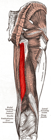
Description
Semitendinosus is one of the three muscles that make up the hamstrings muscle group, and it is located at the posterior and medial aspect of the thigh. The semitendinosus is so named due to it having a long tendon of insertion.
The muscle is fusiform and ends a little below the middle of the thigh in a long round tendon which lies along the medial side of the popliteal fossa; it then curves around the medial condyle of the tibia and passes over the medial collateral ligament of the knee-joint, from which it is separated by a bursa, and is inserted into the upper part of the medial surface of the body of the tibia, nearly as far forward as its anterior crest.
The semitendinosus is more superficial than the semimembranosus (with which it shares very close insertion and attachment points). However, because the semimembranosus is wider and flatter than the semitendinosus, it is still possible to palpate the semimembranosus directly.
At its insertion it gives off from its lower border a prolongation to the deep fascia of the leg and lies behind the tendon of the sartorius, and below that of the gracilis, to which it is united. These three tendons form the Pes Anserinus, thus named due to the appearance resembling a webbed “goose’s foot”.
Anatomy
Origin
It arises, by a common tendon origin with the long head of the biceps femoris, from from the lower medial facet of the lateral section of the ischial tuberosity. The semitendinosus muscle mainly originates from the medial surface of the tendon of the long head of the biceps femoris, and also originates from the ischial tuberosity with a thin tendon and a muscular part.[1]
Insertion
The semitendinosus tendon inserts at the upper part of the medial surface of the tibia, behind the attachment of sartorius and infero-anterior to the attachment of gracilis.
Nerve Supply
Tibial portion of the sciatic nerve (L5, S1, 2).
Artery
Branches from the internal iliac, popliteal, and profunda femoris arteries.
Function
- Extension of the thigh at the hip
- Agonists: gluteus maximus, semimembranosus, biceps femoris (long head), and adductor magnus (posterior part)
- Antagonists: psoas major and iliacus
The semitendinosus is also a weak medial rotator of the hip.
2. Flexion of the leg at the knee
- Agonists: biceps femoris (long head), biceps femoris (short head), and semimembranosus
- Antagonists: vastus lateralis, vastus medialis, vastus intermedius, and rectus femoris
Gracilis, sartorius, popliteus, gastrocnemius, and plantaris assist with flexion of the knee.
3. Internal rotation of the knee when the knee is flexed
- Agonists: popliteus and semimembranosus
- Antagonist: biceps femoris (long head) and biceps femoris (short head)
Sartorius and gracilis assist with internal rotation of knee.
Clinical relevance
A grafted semitendinosus tendon (sometimes combined with gracilis tendon) may be used to replace an unrepairable cruciate or collateral ligament, a service that was previously provided by a graft from the patellar ligament. While either donor site may be used, the hamstring choice provides less post-operative consequences to kneeling.
Assessment
With the patient lying prone, the semitendinosus can be palpated by locating the space between the two large bands that comprise the hamstring tendons just superior to the posterior knee. Palpate medially to this space to locate the semitendinosus tendon and proximal to the tendon for the semitendinosus muscle.
Resources
References
- ↑ Sato K, Nimura A, Yamaguchi K, Akita K. ↑ Dr Nabil Ebraheim. Anatomy Of The Semitendinosus Muscle – Everything You Need To Know. Available from: function gtElInit() { var lib = new google.translate.TranslateService(); lib.setCheckVisibility(false); lib.translatePage('en', 'pt', function (progress, done, error) { if (progress == 100 || done || error) { document.getElementById("gt-dt-spinner").style.display = "none"; } }); }

