Description
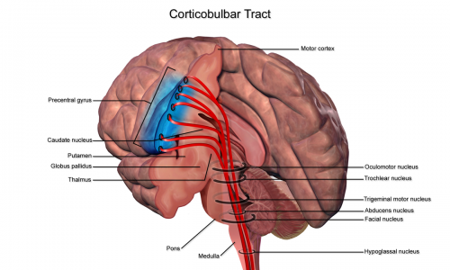
The corticobulbar tract is a descending pathway responsible for innervating several cranial nerves, and runs in paralell with the corticospinal tract[1].
Anatomy
Origin
– Motor cortex (precentral gyrus and anterior part of the paracentral lobule) [1]
Course / Path
– The corticobulbar tracts leave the internal capsule and enter the basilar part of the pons as numerous bundles [1] The fibres leave the cerebral crus adjacent to the corticospinal tract.[1] The fibres can take several paths and have several different terminations:
i) Termine directly on alpha motor neurones or interneurones innervating alpha motorneurons in the brainstem. These control somatic motor acitivity in the head e.g. muscles that control mastication, expression and eye movement.[2]
ii) Axons that innervate motor nerve cranial nuclei can decussate (cross) before they terminate, resulting in them innervating contralateral muscles. As some decussate and some descend ipsilaterally, it results in bilateral descending control. [2]
iii) Directly innervate cranial nervces or through interneurones I.E. via the corticospinal tract. [2]
Function
– Innervates muscles of the face, tongue, jaw, and pharynx, via the cranial nerves.[3]
– The corticobulbar tract directly innervates the nuclei for cranial nerves:
V – Trigeminal- muscles for mastication
VII- Facial- muscles of the face
XI- Accessory- sternocleidomastoid and trapezius
XII- Hypoglossal- muscles of the tongue [4]
– Cranial nerves motor regions of X (vagus nerve) in the nucleus ambiguus.[4]
Pathology
– Unilateral leisons to the corticobular usually do not result in any clinical effect on the neck and head muscles as the lower motor neurons of the brain stem recieve bilateral corticobulbar innervation. [5] However there are two exceptions to these rules:
1) Facial nucleus (VII): The muscles of the lower face recieve contralateral input from the opposite motor cortex. Therefore contralateral leisons to the motor cortex/internal capsule results in weakness to the face muscles in the opposite side of the face. However they would still be able to wrinkle their forehead as this is bilaterally innervated by the corticobulbar tract. [5]
2) Hypoglossal nucleus (XII): The genioglossus muscle (muscle responsible for sticking out the tongue) receives innervation from the contralateral motor cortex. Therefore lesion involving the right motor cortex/ internal capsule would result in weakness in the left hypoglossal muscle. Therefore due to the weakness in the left side of the tongue, the strong muscle on the right side, pushes the tongue to the left. [5]
Resources
References
- ↑ 1.01.11.21.3 Young, Paul. Med school Anatomy. CHAPTER 4. THE PYRAMIDAL SYSTEM. ↑ 2.02.12.2 Brown, AC. Physiology & Neuroscience Web Sites. Neuroscience: Motor Systems. ↑ The Brain from Top to bottom. THE AXONS ENTERING AND LEAVING THE MOTOR CORTEX. ↑ 4.04.1 Revolvy. Corticobulbar tract. ↑ 5.05.15.2 HyperBrain. A resource for learning Neuroanatomy. Chapter 10. Lower and Upper Motor Neurons and the Internal Capsule. ↑ Soton Brain Hub. Corticobulbar tract.

