

 Cart is empty
 O latíssimo do dorso, também chamado de grande dorsal, é um músculo largo e triangular das costas, importante em exercícios como barra fixa e natação.

O latíssimo do dorso, também chamado de grande dorsal, é um músculo largo e triangular das costas, importante em exercícios como barra fixa e natação.
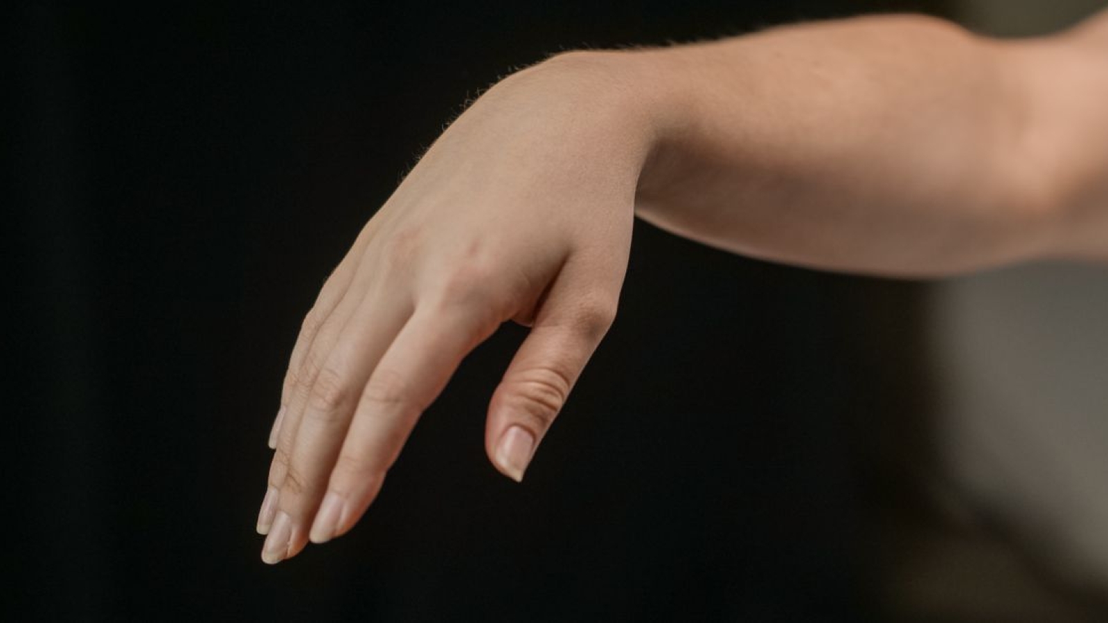
O extensor radial longo do carpo é um músculo do compartimento posterior do antebraço que estende e abduz o punho, além de auxiliar na flexão do cotovelo e na preensão.
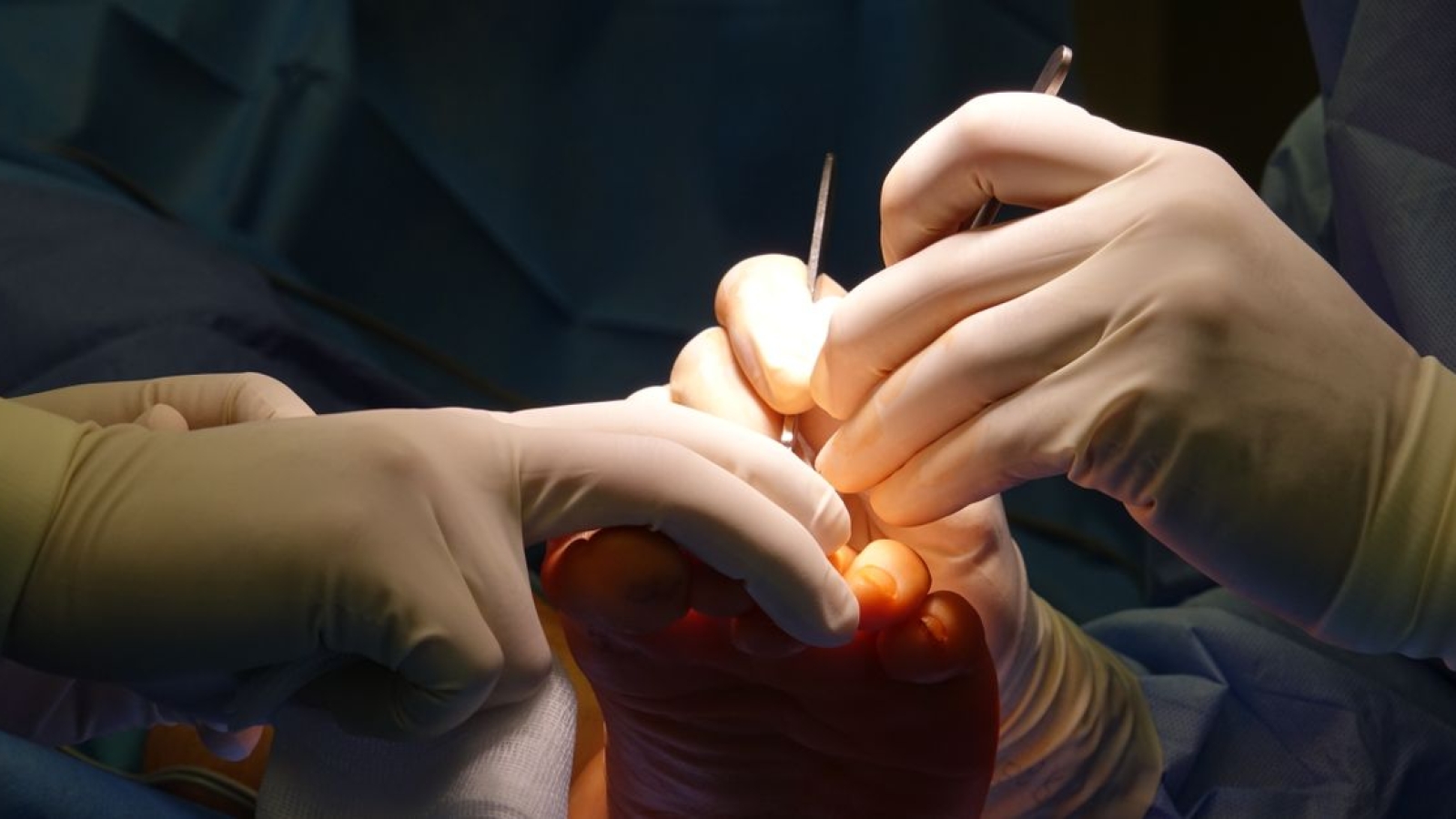
A artéria dorsal do pé (artéria dorsalis pedis) é um vaso sanguíneo do membro inferior que leva sangue oxigenado à superfície dorsal do pé. Origina-se na face anterior da articulação do tornozelo, como continuação da artéria tibial anterior.
A sindesmose tibiofibular distal é uma articulação fibrosa entre tíbia e fíbula, estabilizada por ligamentos e membrana interóssea, essencial para o tornozelo.

A costocondrite é uma inflamação dolorosa das articulações costocondrais, de caráter autolimitado. Saiba diferenciar da síndrome de Tietze e conhecer a anatomia relevante.
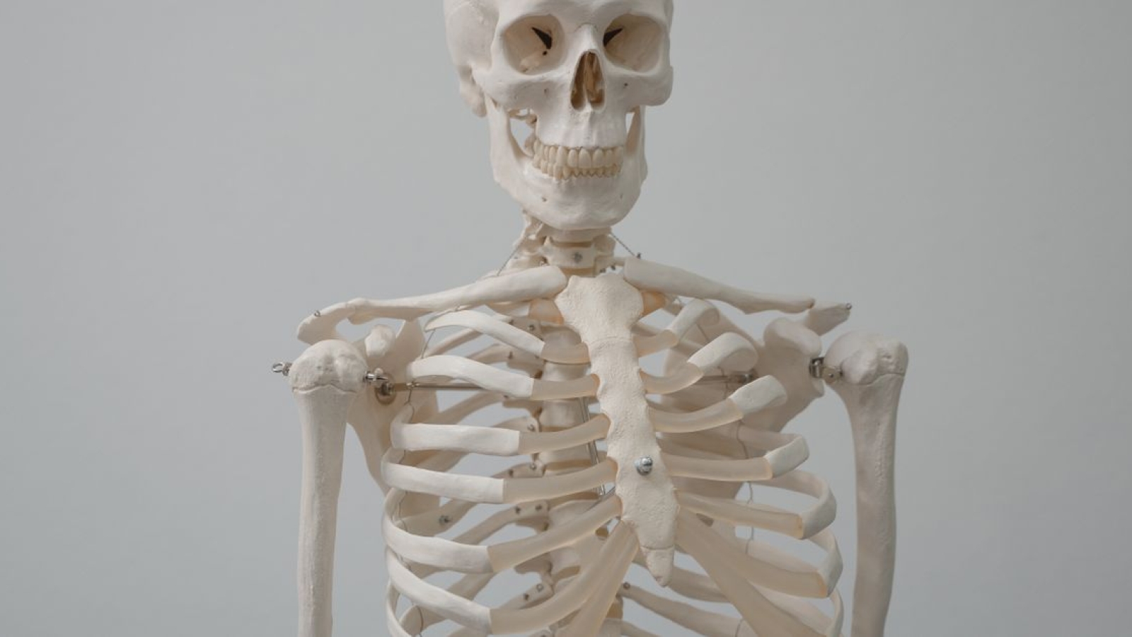
As articulações costovertebrais conectam as costelas às vértebras torácicas, permitindo movimentos de deslizamento. Conheça sua anatomia, ligamentos e função.
O trato corticospinal (TCS) é uma via descendente essencial para o controle motor voluntário, especialmente dos movimentos distais. Este artigo aborda sua anatomia, função e importância clínica.
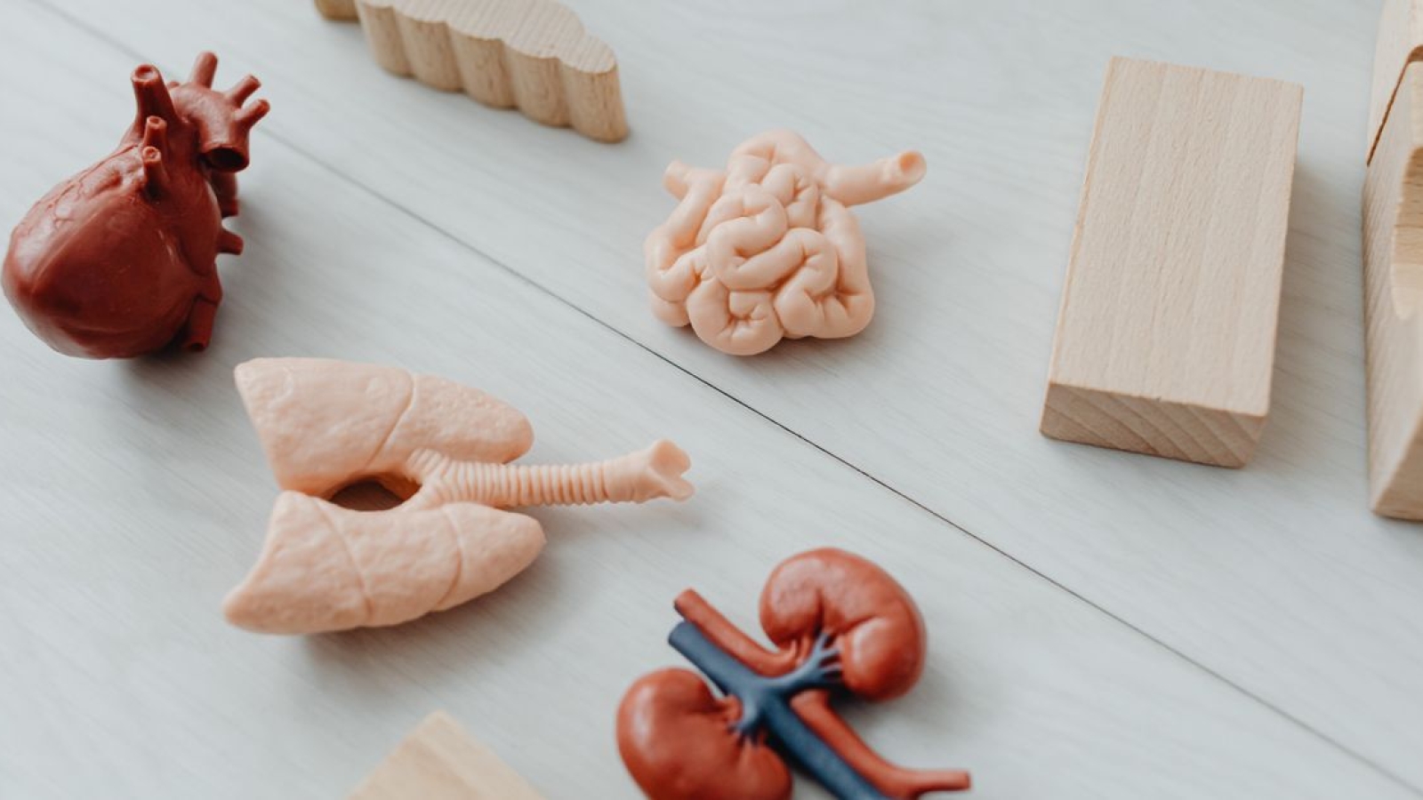

The lungs, which is the organ for respiration is a paired cone shaped organs lying in the thoracic cavity separated from each other by the heart and other structures in the mediastinum.
Each lung has a base resting on the diaphragm and an apex extending superiorly to a point approximately 2.5 cm superior to the clavicle[1]. It also has a medial surface and with three borders- anterior, posterior and inferior. The broad coastal surface of the lungs is pressed against the rib cage, while the smaller mediastinal surface faces medially. The lungs receives the bronchus, blood vessels, lymphatic vessels and nerves through a slit in the mediastinal surface called the helium, and the structures entering the helium constitutes the lungs root[2].
The right lung is larger and weighs more than the left lung[3][2]. Since the heart tilts to the left, the left lung is smaller than the right and has an indentation called the cardiac impression to accommodate the heart. This indentation shapes the inferior and anterior parts of the superior lobe into a thin tongue-like process called the lingual.
Each lung is invested by and enclosed in a serous pleural sac that consists of two continuous membranes.
The visceral or pulmonary pleura invest the lungs,
The parietal pleura line the pulmonary cavities and adhere to the thoracic wall, mediastinum and diaphragm.

The parietal pleura consist of four parts: coastal pleura which lines the internal surface of the thoracic wall, mediastinal pleura which lines the lateral aspect of the mediastinum, diaphragmatic pleura which lines the superior surface of the diaphragm on each side of the mediastinum, cervical pleura extends through the superior thoracic aperture into the root of the neck, forming a cup-shaped dome over the apex of the lung[2].
The pleural cavity is the potential space between the visceral and parietal layers of the pleural and it contains a capillary layer of serous pleural fluid which lubricates the pleural surfaces and allows the layers to slide smoothly over each other during respiration. Surface tension created by the pleural cavity provides the cohesion that keeps the lung surface in contact with the thoracic wall[1].

Each lung is divided into lobes by fissures.
Both lungs have oblique fissure and the right is further divided by a transverse fissure. The oblique fissure in the left lung separates the superior and the inferior lobe. The oblique and horizontal fissure divides the lungs into superior, middle and inferior lobes. Thus the right lung has three lobes while the left has two.
Each lobe is supplied by a lobar bronchus. The lobes are subdivided by bronchopulmonary segments which are supplied by the segmental bronchi.
All the respiratory passages from the trachea to the respiratory bronchioles are called the tracheobronchial tree[1][2][3]. The trachea divides at the sternal angle into right and left primary bronchus which goes into the right and left lungs. Each bronchus enters the lung at a notch called the hilum. Blood vessels and nerves also connect with the lungs here and together with the bronchus forms a region called the root of the lungs.
The right main bronchus is larger in diameter and more vertical making it directly in line with the trachea than the left main bronchus[2]. Thus swallowed objects that accidentally enter the lower respiratory tract are most likely to become lodged in the right main bronchus[4].

The main bronchi divide into lobar or secondary bronchi within each lung. Two lobar bronchi exist in the left lung, and three exist in the right lung. The lobar bronchi, in turn give rise to segmental or tertiary bronchi. The tertiary bronchi supply the bronchopulmonary segments.
Functionally, the lung is divided into a series of bronchopulmonary segments. The bronchopulmonary segments are the largest subdivision of a lobe. They are separated from adjacent segments by connective tissue septa and are also surgically resectable. They are 10 bronchopulmonary segments in the left lung and 8-10 in the left lung[5].
The bronchi further divides, finally giving rise to the bronchioles which are less than 1mm in diameter. Each bronchioles divides into 50 to 80 terminal bronchioles, the final branches of respiratory bronchioles. The functional unit of the lungs which is the acinus includes the respiratory bronchioles, alveolar ducts, and sacs and the alveolar[1]. Approximately 16 generations of branching occur from the trachea to the terminal bronchioles[1]. As the air passageways of the lungs become smaller, the structure of their walls changes[3].
The major airways from the trachea through the ten generations of bronchi have descending amounts of cartilaginous support surrounded by smooth muscles and elastic fibres, they have goblet cells for mucus production and are lined with ciliated columnar epithelium to facilitate secretion clearance.
The next five generation of bronchioles have no cartilage or goblet cells, but still have elastic tissue and smooth muscle fibres. They lined with ciliated cuboidal epithelium. Relaxation and contraction of the smooth muscles of the bronchi and bronchioles can change the diameter of the air passageways and so change the volume of air moving through them. The alveoli provide a large surface area for gas exchange. The alveolar walls consist of simple squamous epithelium. Also the tissues surrounding the alveoli contain elastic fibres which allow the alveoli to expand during inspiration and recoil during expiration.
The epithelium of the respiratory zone is not ciliated but debris from the air can be removed by macrophages that move over the surface of the cells. Approximately 300 million alveoli are in the lungs. The average diameter of the alveoli is approximately 250um and their walls are extremely thin. There are two types of cells in the alveoli: type I pneumocytes which are thin squamous epithelia cells that forms 90% of the alveoli surface. Most of the gas exchange take place between these cells. Type II pneumocytes are round secretory that produce surfactant, which makes it easier for the alveoli to expand during inspiration[5].
The bronchial arteries arising from the aorta provide blood supply to the non-respiratory airways, pleura, and connective tissue while the pulmonary arteries supply the respiratory units (acini) and participate in gas exchange.
Venous drainage is mainly by the pulmonary veins (right and left superior and inferior pulmonary veins), though the venous of drainage from the walls of the larger bronchi is carried out by the bronchial veins. All four veins (pulmonary veins) drain into the left atrium.
The lungs and airways are innervated by the branches of sympathetic trunk and vagus nerve[2]. Sympathetic nervous stimulation results in bronchodilation and slight vasoconstriction, while parasympathetic nervous system stimulation results in bronchoconstriction and indirect vasodilation. The function of the lungs is controlled through the respiratory centre with groups of neurons located at the pons and the medulla oblongata, and complex interactions of specialized peripheral central chemoreceptors.
Auscultation of the lungs and percussion of the thorax are important techniques used during physical examination. Auscultation assesses airflow through the tracheobronchial tree into the lungs.
Percussion helps establish whether the underlining tissues are air filled (resonant sound), fluid filled (dull sound) or solid (flat sound).
Based on the anatomy of the bronchial tree, gravity can be used to assist the clearance of bronchial secretions through the use of positioning. Also Bronchial disorders such as tumour and abscesses may be localized in a bronchopulmonary segments and may be surgically removed without disrupting the surrounding lung tissue.
There are a number of structures that protects the lungs from inhaled organisms and particles:

O Shiatsu combina um movimento suave da energia do ki com os benefícios físicos de uma massagem profunda nos tecidos. Além disso, combina o conhecimento ocidental da anatomia e os fundamentos fisiológicos e filosóficos da fisiologia da Medicina Tradicional Chinesa (MTC) .

O Shiatsu é realizado em uma cama de massagem baixa ou, alternativamente, no chão. Ao contrário de outras terapias de massagem, nenhum óleo é aplicado para que você permaneça totalmente vestido durante todo o tratamento e, portanto, é aconselhável que você use roupas confortáveis. A terapia não é dolorosa, mas sim uma sequência rítmica contínua, usando dedos, polegares e palmas para aplicar pressão. Alguns pontos de pressão podem parecer mais suaves, mas depois você se sentirá aliviado.
Desenvolvido no Japão, o Shiatsu usa a pressão do dedo em pontos específicos do corpo, movimentos de balanço, alongamentos e rotações articulares para restaurar o fluxo saudável de energia ( chi em chinês, ki em japonês) para o corpo. O Shiatsu é holístico, abordando todo o corpo, em vez de se concentrar em uma área onde os sintomas são mais óbvios.
O nome do shiatsu vem de duas palavras japonesas – shi (dedo) e atsu (pressão) – mas um praticante também pode aplicar pressão usando outras partes da mão, cotovelos e joelhos. Nenhum óleo é usado neste tratamento.

A técnica foi formalmente nomeado no início do século 20, mas tem raízes na Medicina Tradicional Chinesa (MTC). A teoria por trás do shiatsu, como a acupuntura, é que o corpo tem caminhos de energia invisíveis, ou meridianos, ao longo dos quais a energia do corpo flui.
Quando você está saudável, a energia flui livremente ao longo dos meridianos, suprindo todas as partes do corpo com energia vital. Mas quando o corpo foi enfraquecido por uma dieta pobre, cafeína, drogas, álcool e estresse emocional, o kinão flui mais suavemente. Pode ser deficiente em algumas áreas e excessivo em outras.

O praticante conhece esses caminhos de energia, bem como os pontos (chamados tsuobos em japonês) que estão localizados ao longo dos meridianos. São essencialmente áreas de alta condutividade e podem ser afetadas por diversas modalidades: pressão dos dedos no shiatsu; agulhas em acupuntura; calor na moxabustão.
Curso de Ventosas Terapeuticas
Ao aplicar pressão a esses tsuobos , o praticante identifica bloqueios e desequilíbrios e faz com que a energia flua suavemente novamente. Se a energia ou ki é deficiente, o praticante introduz energia nessa área com seu toque. Se o ponto for difícil e dolorido ao toque, há um excesso de ki que o praticante precisa drenar. Como acontece com qualquer tratamento, você controla a quantidade de pressão que deseja. Uma sessão de shiatsu geralmente dura entre 45 minutos e uma hora.
 Tornando isso um pouco mais complicado para a mente ocidental é que cada caminho de energia está relacionado a um órgão (rins, pulmões, fígado, coração, estômago etc.) assim como uma emoção ou estado mental (medo, tristeza, raiva). É interessante, mas você não precisa se preocupar com isso. Se houver sensibilidade no meridiano do seu fígado, isso não significa que você tem doença hepática. Significa apenas que a energia do seu fígado está desequilibrada.
O modelo oriental tradicional de saúde e bem – estar é muito diferente do modelo ocidental e é mais uma questão de restaurar a saúde e o equilíbrio do corpo antes que algo de errado. É também sobre preservar o seu ki , que fica mais fraco com a idade.
Se você tem câncer de mama e está interessado em encontrar um praticante de Shiatsu, peça recomendações ao seu cirurgião ou oncologista. É importante que sua equipe de saúde saiba que você está considerando o Shiatsu. Também é importante informar ao praticante sobre seu diagnóstico, tratamento e qualquer sintoma que você possa ter.
Se você foi diagnosticado com câncer de mama, também tenha em mente:

A subluxação ou luxação dos tendões peroneais é um distúrbio envolvendo um alongamento, uma ruptura ou uma avulsão do retináculo do fibular superior [1] . Também pode haver subluxação dos tendões com um SPR intacto (subluxação intramedida) [2] .
O músculo fibular curto e o músculo fibular longo estão contidos no sulco retromaleolar da fíbula. A profundidade do sulco é variável e foi observada como ausente ou convexa. [3] Os tendões são estabilizados por um retináculo peroneal superior. O SPR é formado pelo espessamento da aponeurose superficial. Uma pequena crista fibrosa é ocasionalmente vista originando-se da fíbula distal próxima à origem da SPR e aumenta a profundidade do sulco fibular. Distal à fíbula está o retináculo fibular inferior, que cobre os tendões por cerca de 2 a 3 cm distalmente à ponta da fíbula [4] .
O mecanismo mais comum é uma força de dorsiflexão no tornozelo associada a uma contração rápida e forte dos tendões peroneais e com uma eversão do retropé. Os tendões fibulares longos e brevis subluxam ou deslocam-se do sulco retro-retroolar lateral. Isso resulta de uma ruptura ou avulsão ou frouxidão significativa do SPR. Alguns pacientes apresentam uma apresentação mais crônica e não conseguem lembrar de um episódio traumático. Também são relatados fatores congênitos, por exemplo, o sulco ou a crista que ajuda a aprofundar o sulco pode ser muito superficial ou mesmo ausente ou a SPR pode ser muito solta [5] . Além dos fatores congênitos, o mecanismo mais comum é uma força dorsiflexória no tornozelo associada à contração forçada concomitante dos tendões fibulares combinados com a eversão do retropé posterior.[5] .
Subluxação do tendão fibular é comumente encontrada no esqui, mas também tem sido relatada em outros esportes [1] [6] .

Existem três graus para classificar a subluxação peroneal aguda [7] [3] [8]
Grau I: O retináculo, que é confluente com o periósteo na fíbula, é retirado da fíbula, resultando em luxação dos tendões.
Grau II: A crista fibrocartilaginosa e a SPR são avulsionadas do aspecto posterior da fíbula.
Grau III: Avulsão óssea do aspecto posterolateral da fíbula contendo a borda cartilaginosa e um floco de osso, permitindo que o tendão deslize sob o periósteo.
Mais tarde, Oden descreveu um quarto ano:
Grau IV: O SPR elevado do calcâneo
A subluxação peroneal aguda é freqüentemente difícil de ser avaliada clinicamente. Geralmente há edema, equimose e sensibilidade pontual na mesma distribuição do que no caso de entorse de tornozelo, dificultando o diagnóstico [5] .
Em situações crônicas, os pacientes freqüentemente se queixam de um mecanismo doloroso do vento e podem tipicamente reproduzir a luxação por eversão ativa da dorsiflexão do pé [5] . Na maioria dos casos, um tendão peroneal que se estica ao redor da margem posterior do maléolo lateral pode ser palpado e até visualizado pelo examinador. Esses achados clínicos podem ser melhorados pela aplicação de uma leve resistência à eversão da exérese dorsifl.
 Em recém-nascidos e recém-nascidos, o tratamento conservador do tendão fibular subluxar é a regra e a resolução espontânea é extremamente alta [5] , [9] . Nos adultos, o tratamento pode ser dividido em não-operatório e operatório, quando se considera lesões agudas ou crônicas [5] . Quando uma subluxação aguda é diagnosticada, o paciente pode considerar tratamento não cirúrgico [7] [3] . Se o tratamento conservador falhou ou se há uma subluxação crônica, o tratamento cirúrgico é provavelmente necessário [7] . O tratamento precoce é crítico, pois é mais provável que um tendão que continua a subluxar (sair da posição) rasgue ou rompa [10] .
Em particular, o tratamento não cirúrgico é indicado para lesões grau I e possivelmente grau III, dependendo do grau de deslocamento do fragmento cortical [7] . O tratamento conservador está associado a riscos mínimos, mas também a uma alta taxa de falha [5] .
O tratamento não operatório consiste na aplicação de um molde sem suporte de peso por 4 a 6 semanas [7] . Esta gestão conservadora visa permitir que o SPR volte a aderir ao aspecto posterolateral da fíbula [5] .
Tentativas conservadoras também podem incluir cintas adesivas com uma almofada para restringir a subluxação, almofadas em forma de J que ancoram na frente da fíbula e envolvem lateralmente e posteriormente para manter os tendões no lugar e não suportar o peso por 4-6 semanas. Se o pé for mantido relativamente estável e a fita restringir o movimento do tendão, a formação de tecido cicatricial pode permitir que o tendão seja manuseado sem cirurgia [11] .
Menos controvérsia existe para o manejo das lesões crônicas, pois o reparo cirúrgico da subluxação do tendão peroneal fornece bons resultados na maioria dos casos [5] .
Opções cirúrgicas [7] [12] :
A abordagem cirúrgica pode diferir com base no grau de lesão [4] .
Com exceção das precauções de movimento, a reabilitação pode prosseguir de maneira semelhante à entorse crônica do tornozelo. No entanto, a reabilitação após a cirurgia provavelmente levará mais tempo para restaurar totalmente o movimento, a força e a função [12].
Após a cirurgia, o tornozelo é mantido por 4 semanas em um molde sem suporte de peso, seguido por 2 semanas em um molde de suporte de peso. Durante a imobilização, o condicionamento cardiovascular é realizado juntamente com o fortalecimento muscular proximal [12] .
Os primeiros tratamentos de fisioterapia são projetados para ajudar a controlar a dor e inchaço da cirurgia. Tratamentos com gelo e estimulação elétrica podem ser usados. O terapeuta também pode usar massagens e outros tratamentos manuais para aliviar o espasmo muscular e a dor. A mobilização de tecidos moles ao redor do local da cicatriz pode ser empregada para aumentar a mobilidade dos tecidos moles [11] .
Os tratamentos também são usados para ajudar a melhorar a amplitude de movimento do tornozelo por resistência progressiva e exercícios de ROM, sem colocar muita pressão sobre a área [12] . Dorsiflexão ativa e resistida e eversão são evitadas durante a fase inicial de reabilitação para reduzir o estresse na SPR (aproximadamente 6 a 8 semanas [11] .
Exercícios de mobilização talar e dorsiflexão ativa e eversão iniciam quando o paciente pode suportar peso sem dor [11] .
A progressão dos exercícios resistidos de fortalecimento, propriocepção e agilidade é iniciada quando o paciente pode suportar peso sem dor e sem apoio. À medida que a força e a propriocepção melhoram, o paciente pode progredir através de atividades pliométricas e funcionais que levam a um retorno à competição [11] .