Acupuntura o que é para que serve e como funcional

Acupuntura o que é para que serve e como funcional

Qual é o significado de acupuntura?

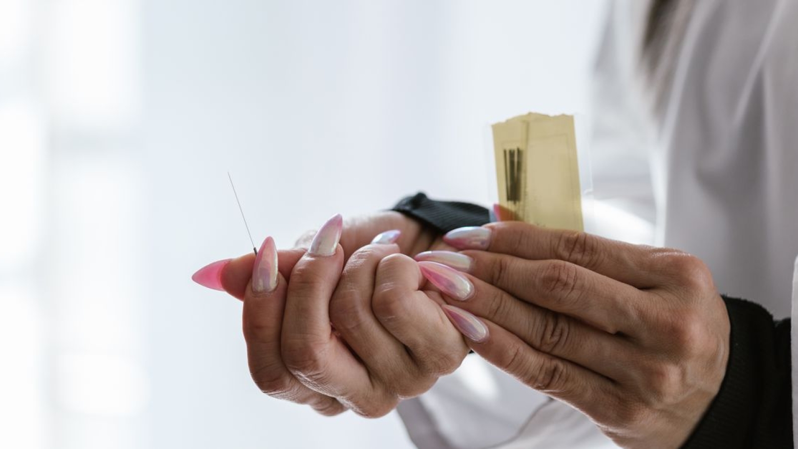
Acupuntura é uma terapia alternativa que se originou na China há mais de 2.500 anos. É uma forma de medicina tradicional que utiliza agulhas finas e leves para estimular pontos específicos no corpo, conhecidos como pontos de acupuntura. O objetivo da acupuntura é equilibrar o fluxo de energia, ou “Qi” (pronunciado como “chee”), que percorre o corpo através de canais, ou “meridianos”.

Na medicina tradicional chinesa, o Qi é visto como o responsável pelo bem-estar e saúde do corpo. Se o Qi estiver bloqueado ou desequilibrado, pode levar a problemas de saúde, incluindo dor, ansiedade, insônia, depressão, entre outros. A acupuntura visa ajudar a restaurar o equilíbrio do Qi e promover a cura natural, ajudando a melhorar a saúde e bem-estar geral.

A acupuntura é uma forma de tratamento não invasiva e segura e é amplamente utilizada em todo o mundo para tratar uma ampla gama de condições, incluindo dor crônica, dor de cabeça, enxaqueca, dor nas costas, dor muscular, insônia, ansiedade, depressão, entre outros. Além disso, a acupuntura é frequentemente combinada com outras formas de terapia, como massagem, exercícios físicos e alimentação saudável, para promover uma abordagem mais holística e integral para a saúde e bem-estar.

Qual é o significado de acupuntura?

O que a acupuntura pode fazer por você?

Pode ajudar a tratar uma ampla gama de condições e problemas de saúde, incluindo:

  1. Dor crônica: É eficaz para aliviar a dor crônica, incluindo dor nas costas, dor lombar, dor no pescoço, dor de cabeça, dor menstrual, entre outros.
  2. Ansiedade e estresse: A acupuntura pode ajudar a reduzir a ansiedade e o estresse, melhorando o bem-estar emocional e a qualidade de vida.
  3. Insônia: É eficaz para tratar a insônia e ajudar a melhorar a qualidade do sono.
  4. Depressão:  Pode ser útil no tratamento da depressão, ajudando a melhorar o humor e a reduzir os sintomas.
  5. Enxaqueca e dor de cabeça: A acupuntura é eficaz para tratar enxaqueca e dor de cabeça, ajudando a reduzir a frequência e intensidade dos ataques.
  6. Síndrome do Intestino Irritável (SII): A acupuntura pode ajudar a aliviar os sintomas da SII, incluindo dor abdominal, diarreia, constipação, entre outros.
  7. Problemas de saúde feminina: A acupuntura pode ser útil no tratamento de problemas de saúde feminina, incluindo dismenorreia, síndrome pré-menstrual (TPM), menopausa, entre outros.
  8. Outras condições: A acupuntura também pode ser útil no tratamento de outras condições, incluindo asma, doenças cardíacas, fadiga, síndrome do túnel do carpo, entre outros.

Em geral, a acupuntura é uma forma segura e eficaz de tratar uma ampla gama de condições e problemas de saúde. Além disso, a acupuntura pode ser combinada com outras formas de terapia, como massagem, exercícios físicos e alimentação saudável, para promover uma abordagem mais holística e integral para a saúde e bem-estar.

A acupuntura é uma técnica milenar de medicina tradicional chinesa que foi criada há mais de 2.500 anos. Ela se baseia na filosofia de que existe um fluxo de energia vital, conhecido como “Qi”, que circula pelo corpo humano através de meridianos ou canais específicos. Quando o equilíbrio deste fluxo de energia é perturbado, isto pode levar a uma variedade de condições de saúde, incluindo dor, doenças e desordens emocionais.

A acupuntura é realizada inserindo agulhas muito finas em pontos específicos ao longo dos meridianos do corpo, a fim de restaurar o equilíbrio do fluxo de energia e promover a cura natural. Além disso, a acupuntura também pode incluir a estimulação de pontos de pressão, moxabustão (queimadura de ervas medicinais), ou outras técnicas complementares.

A acupuntura é utilizada para tratar uma ampla variedade de condições de saúde, incluindo dor crônica, enxaqueca, insônia, ansiedade, depressão, problemas digestivos, síndrome da fadiga crônica, entre outros. Além disso, a acupuntura também é utilizada como uma forma complementar de tratamento para doenças graves, tais como câncer e doenças cardiovasculares.

Em geral, é considerada uma terapia segura e eficaz, com poucos efeitos colaterais. No entanto, é importante procurar um acupunturista qualificado e licenciado para garantir que a terapia seja realizada de forma adequada e segura. Além disso, é importante lembrar que a acupuntura não deve ser utilizada como substituto para o tratamento convencional recomendado por um médico, mas sim como uma forma complementar de cuidado de saúde.

Você sabe a diferença entre a acupuntura e a auriculoterapia

A acupuntura e a auriculoterapia são duas técnicas de medicina alternativa baseadas na filosofia da medicina tradicional chinesa. Embora compartilhem algumas similaridades, existem algumas diferenças importantes entre elas.

A acupuntura se concentra na inserção de agulhas em pontos específicos ao longo dos meridianos do corpo para restaurar o equilíbrio do fluxo de energia e promover a cura natural. Por outro lado, a auriculoterapia se concentra na estimulação dos pontos de acupuntura na orelha externa, que são considerados reflexos dos órgãos e sistemas do corpo.

A auriculoterapia é geralmente considerada uma forma menos invasiva de acupuntura e é especialmente útil para tratar condições relacionadas ao sistema nervoso central e ao sistema endócrino. Além disso, a auriculoterapia também é utilizada para tratar uma ampla gama de outras condições de saúde, incluindo dor crônica, problemas de sono, ansiedade, depressão, entre outros.

Em resumo, a acupuntura e a auriculoterapia são duas técnicas complementares, cada uma com suas próprias aplicações e vantagens. É importante consultar um profissional de saúde qualificado para determinar qual técnica é a mais adequada para cada indivíduo e condição de saúde.

A acupuntura doi?

A acupuntura geralmente não é dolorosa, e muitas pessoas descrevem a sensação como um leve formigamento ou pressão. Algumas pessoas podem sentir uma sensação de picada no momento da inserção da agulha, mas isso é geralmente breve e se dissipa rapidamente.

O grau de sensibilidade varia de pessoa para pessoa, e algumas pessoas podem ser mais sensíveis ao toque ou à estimulação do que outras. No entanto, os acupunturistas são treinados para inserir as agulhas de maneira segura e confortável, e muitas vezes podem usar técnicas para minimizar a dor.

Se você tiver medo de agulhas ou é sensível ao toque, é importante discutir suas preocupações com seu acupunturista antes de iniciar a terapia. Eles podem trabalhar com você para encontrar a melhor maneira de tornar a terapia confortável e eficaz para você.

E o que é acupuntura sistemica? para que ela serve?

A acupuntura sistêmica é uma abordagem de acupuntura que se concentra na avaliação e tratamento dos sistemas corporais como um todo, em vez de apenas tratar uma condição específica. Em vez de se concentrar em um único sintoma ou problema, a acupuntura sistêmica procura compreender como o corpo como um todo está funcionando e onde há desequilíbrios ou bloqueios de energia.

Ela baseada na filosofia da medicina tradicional chinesa, que acredita que todos os aspectos da saúde mental, emocional e física estão interconectados. Por esse motivo, a acupuntura sistêmica pode ser uma abordagem eficaz para tratar uma ampla gama de condições, desde problemas de saúde comuns como dor de cabeça, dor nas costas e dor muscular, até condições mais complexas, como ansiedade, insônia, depressão, entre outras.

A acupuntura sistêmica pode ser especialmente útil para pessoas que estão procurando uma abordagem mais holística e integral para a saúde e bem-estar. Ao trabalhar com o corpo como um todo, é possível restaurar o equilíbrio do fluxo de energia e promover a cura natural, ajudando a melhorar a saúde e bem-estar geral.

Pode realizar acupuntura no pé?

Sim, é possível realizar. A auriculoterapia, ou acupuntura auricular, é uma forma de acupuntura que se concentra na orelha e é muito eficaz para tratar uma ampla gama de condições, incluindo dor, ansiedade, insônia, estresse, entre outros.

A auriculoterapia é baseada na ideia de que a orelha é uma representação miniatura do corpo humano, com cada ponto na orelha correspondendo a uma parte específica do corpo. Ao estimular esses pontos, é possível equilibrar o fluxo de energia e promover a cura natural.

Além da auriculoterapia, pode ser realizada em outras áreas do pé, incluindo o arco do pé e as solas dos pés. Essas áreas são ricas em terminações nervosas e são consideradas importantes para a circulação de energia e a saúde geral do corpo.

A acupuntura no pé é uma forma eficaz de tratar uma ampla gama de condições, incluindo dor, ansiedade, insônia, estresse, entre outros, e pode ser uma opção eficaz para pessoas que buscam uma abordagem mais holística e integral para a saúde e bem-estar.

Você deve gostar tambem

Conheça sobre o Manguito Rotador e suas Funções

Curso de Auriculoterapia

Auriculoterapia para que Serve

Pós-graduação em Ortopedia com ênfase em Terapia Manual

Que roupa usar na sessão de acupuntura?

Nas sessões de acupuntura, é recomendável vestir roupas confortáveis e fáceis de remover, especialmente se a acupuntura for realizada nas pernas, braços ou costas. Alguns acupunturistas podem precisar acessar áreas do corpo que exigem a remoção de roupas, por isso é importante levar em consideração o conforto durante a sessão.

No geral, roupas soltas e leves são as melhores, pois permitem que o acupunturista acesse facilmente as áreas do corpo que precisam ser tratadas. Se a acupuntura for realizada no abdômen ou na região lombar, é possível que seja necessário remover a roupa superior, por isso é importante levar em consideração essas questões ao escolher as roupas para a sessão.

Em geral, o objetivo é se sentir o mais confortável possível durante a sessão, para que você possa se concentrar nas sensações e obter o máximo de benefícios da acupuntura.

Por que acupuntura relaxa?

A acupuntura pode relaxar o corpo e a mente de várias maneiras. Aqui estão algumas das razões pelas quais a acupuntura pode ser relaxante:

  1. Estimulação dos pontos: a estimulação dos pontos de acupuntura através da inserção de agulhas pode estimular a liberação de endorfinas e outras substâncias químicas no corpo que são conhecidas por ajudar a aliviar a dor e reduzir o estresse.
  2. Relaxamento muscular: pode ajudar a aliviar a tensão muscular, o que pode ajudar a relaxar o corpo e a mente.
  3. Redução do estresse: a acupuntura pode ajudar a reduzir o estresse e a ansiedade, o que pode ter um efeito positivo sobre o bem-estar geral e a capacidade de relaxar.
  4. Estimulação do sistema nervoso parassimpático: a acupuntura pode estimular o sistema nervoso parassimpático, que é responsável por regular o equilíbrio entre o sistema nervoso simpático e o sistema nervoso parassimpático. Quando o sistema nervoso parassimpático é estimulado, pode ajudar a reduzir o estresse e promover o relaxamento.
  5. Efeito placebo: a acupuntura pode ter um efeito placebo positivo, o que significa que o efeito benéfico pode ser atribuído ao poder da sugestão e da crença no tratamento.

Em resumo, pode ser relaxante devido à combinação de estímulo dos pontos de acupuntura, relaxamento muscular, redução do estresse, estimulação do sistema nervoso parassimpático e efeito placebo.

Conteúdos relacionados

O efeito de exercícios de estabilização lombar e mobilização torácica e exercícios em pacientes com dor lombar crônica .

O efeito de exercícios de estabilização lombar e mobilização torácica e exercícios em pacientes com dor lombar crônica .

 
Abstrato

[Objetivo] Investigar se a dor, o equilíbrio e a estabilização da região lombar podem ser melhorados através da mobilização torácica, além de exercícios de estabilização lombar. [Sujeitos e métodos] Este estudo recrutou 36 indivíduos com dor lombar crônica com duração superior a 12 semanas. Os sujeitos recrutados para este estudo participaram voluntariamente e deram seu consentimento assinado à participação. [Resultados] A melhora no equilíbrio foi maior no grupo de exercícios de estabilização lombar, seguido pelo grupo de mobilização e exercícios torácicos e o grupo de fisioterapia tradicional, em ordem decrescente de efeito. [Conclusão] Em conclusão, os exercícios de estabilização lombar combinados à mobilização torácica tiveram maiores efeitos na estabilização do alívio da dor na região lombar,

Palavras-chave: Exercícios de estabilização lombar, Exercícios de mobilização torácica, Dor lombar crônica

INTRODUÇÃO

A dor lombar é uma das doenças mais comuns e cerca de 70 a 80% da população a experimenta uma ou mais vezes na sociedade moderna 1 ) , e sua incidência e os gastos sociais relacionados a ela estão aumentando 2 ) . Existem várias causas de dor nas costas e, entre elas, hérnia de discos intervertebrais e fraqueza muscular causada por dano muscular são as principais causas de dor lombar 3 ) .

A dor lombar é causada pela perda da estabilização segmentar e da capacidade de equilíbrio e limita a amplitude de movimento 4 ) . Os pacientes são designados como tendo dor lombar crônica, quando a dor lombar com causas inespecíficas dura mais de 12 semanas 5 ) . Nos últimos anos, as causas mecânicas da dor são responsáveis ​​pelo maior número de casos. Após analisar o movimento de cada segmento da coluna vertebral de pacientes com disco degenerado, nosso grupo de estudo relatou que a causa mais comum de dor nas costas é a instabilidade da coluna vertebral 6 ) .

No movimento humano em um modelo de múltiplos componentes, o movimento do segmento mais flexível gera o maior ângulo. Portanto, a maioria das disfunções espinhais é causada pela flexibilidade excessiva de um segmento em particular, e não pela flexibilidade 7 ) .

Ao realizar atividades funcionais, a mobilização das articulações adjacentes a um segmento instável é relativamente aumentada, e a diminuição da hipermobilidade por meio da estabilização do segmento hipermóvel pode promover a cicatrização 8 ) . Assim, a mobilização torácica pode ajudar a promover a estabilização da região lombar.

Segundo estudo recente, a instabilidade da coluna lombar é a causa mais importante de lombalgia crônica 9 ) . Se essa instabilidade aumentar, pode provocar reações posturais anormais no equilíbrio estático e resultar na incapacidade de realizar atividades normais devido à perda do equilíbrio sensorial em pacientes com dor lombar crônica. A perda de equilíbrio também pode ser causada pela perda da força muscular ou desequilíbrios neurológicos.

Muitos estudos relataram que os exercícios de estabilização lombar proporcionam estabilização, fortalecendo os músculos profundos lombares de pacientes com dor lombar crônica, mas poucos estudos investigaram a estabilização mecânica lombar por mobilização torácica. Portanto, este estudo investigou se a dor, o equilíbrio e a estabilização da lombar podem ser melhorados através da mobilização torácica, além de exercícios de estabilização lombar.

Estabilização Lombar


ASSUNTOS E MÉTODOS

Este estudo recrutou indivíduos com dor lombar crônica com duração superior a 12 semanas. Quarenta e cinco sujeitos preencheram os critérios de seleção e 36 deles participaram do estudo. Eles foram divididos em 3 grupos; um grupo tradicional de fisioterapia (TPG), um grupo de exercícios de estabilização lombar (LSEG) e um grupo de exercícios de mobilização torácica (TMEG).

Fisioterapia tradicional, exercícios de estabilização lombar ou mobilização e exercícios torácicos foram realizados três vezes por semana, durante 12 semanas. As medidas de desfecho foram uma avaliação visual em escala analógica da dor, o índice de equilíbrio e o comprimento da tradução lombar. Os critérios de seleção foram: experiência de lombalgia ao longo de 12 semanas e participantes que não conseguiam sentar muito tempo em uma posição, que sentiam dor toda vez que mudavam de posição e também frequentemente mudavam sua postura sentada. Os sujeitos escolhidos para este estudo participaram voluntariamente e assinaram o termo de consentimento livre e esclarecido, de acordo com os princípios éticos da Declaração de Helsinque. Todos os indivíduos receberam fisioterapia tradicional, composta por tratamento com compressa quente por 20 minutos, terapia por ultrassom por 5 minutos,

O grupo de exercícios de estabilização lombar, realizou adicionalmente exercícios para estabilizar os músculos multifidus e o transverso abdominal, após receber fisioterapia tradicional. O programa de exercícios de estabilização lombar consistiu em três exercícios. Cada exercício foi repetido 12 vezes em 1 série, e 5 séries, com um intervalo de 3 minutos entre as séries, foram realizadas em uma sessão de terapia. Os três exercícios foram: semi-abdominais, segurando as pernas na posição de pernas cruzadas, com o rosto voltado para baixo e de lado, mantendo a perna levantada.

O grupo de mobilização torácica e exercício realizou adicionalmente dois programas de exercícios e recebeu mobilização passiva de tecidos moles torácicos para aumentar a mobilidade torácica após a fisioterapia tradicional. O programa de exercícios de mobilidade torácica consistiu em 2 exercícios. Cada exercício foi repetido 12 vezes em 1 série, e um total de 5 séries, com um intervalo de três minutos entre as séries, foram realizadas em uma sessão de terapia.

A mobilização de tecidos moles torácicos foi realizada por 5 minutos após a realização dos exercícios de tecidos moles torácicos. Isso envolveu mover a parte superior do corpo, colocar apenas um travesseiro na área torácica, mover a área torácica e mobilizar com a parte superior do corpo e a parte inferior do corpo cruzadas. Os níveis de dor foram avaliados usando uma escala visual analógica (EVA). Neste estudo, o equilíbrio foi avaliado como o melhor escore de três ensaios em uma postura em pé em um instrumento de medição da balança (MICROSWIMG 5.0, HAIDER BIOSWING, Alemanha) conectado a um computador usando o software Posturomed.

Confira tambem

Curso de Estabilização Segmentar

Artigo Sobre Osteopatia

Teste de Yeoman


O teste do comprimento da translação lombar é um teste objetivo da instabilidade lombar. Se qualquer segmento da coluna funcional estiver em um estado instável durante a medição da translação da coluna funcional sob uma força física, ele traduzirá mais do que um segmento estável nas forças físicas da época. No caso de o deslocamento do plano sagital ter sido transferido mais de pelo menos 3 mm em qualquer segmento da curva ou expansão ao realizar a inspeção por raios-X, chamamos de instabilidade da lombar quando mais de 9% do diâmetro do corpo vertebral adjacente foi transferido 10 ) .

O tempo de translação dos segmentos instáveis ​​foi medido da mesma maneira antes e após a intervenção para testar a distância de translação dos segmentos dos pacientes com lombalgia crônica. Para medir a distância, foi traçada uma linha na superfície superior das vértebras inferiores do segmento a ser medida no plano sagital do raio X, juntamente com uma linha vertical. Em seguida, a superfície inferior das vértebras superiores e a parte final da placa final da superfície superior das vértebras inferiores foram conectadas verticalmente a essa linha, e a distância entre as duas linhas foi medida.

Todos os dados obtidos neste estudo foram processados ​​no SPSS para 20.0 Windows. Após a intervenção, as diferenças de estabilidade lombar, equilíbrio e dor entre os grupos foram examinadas pelo teste t pareado. O nível de significância estatística α utilizado foi de 0,05.

RESULTADOS

As características gerais do sujeito são mostradas na Tabela 1 .

Tabela 1.

As características gerais dos sujeitos
Variáveis TPG (N = 12) LSEG (N = 12) TMEG (N = 12)
Duração da dor lombar (anos) 3.3±1.1 2.8±2.2 2.9±1.3
Género masculino (%) 11 (91.7) 10 (83.3) 8 (66.7)
Anos de idade) 45.4±6.4 43.0±6.3 43.0±5.4

Os valores de estabilidade lombar medidos em raios-X são mostrados na Tabela 2 . O grupo de mobilização torácica e exercício e o grupo de exercício de estabilização lombar mostraram melhorias significativas no teste de estabilização lombar.

Mesa 2.

Comparação da estabilização lombar, índice de equilíbrio, dor entre os três grupos
Grupo Estabilização lombar (N = 12) Unidade (mm) Índice de saldo (N = 12) Unidade (pontuação) Dor (N = 12) Unidade (escore)


para postar para postar para postar
lseg 3.7±0.6 1.2±0.5* 179.7±62.3 597.5±40.4* 6.9±0.6 3.1±0.3*
TMEG 3.7±0.7 1.54±0.5* 167.9±47.6 450.6±50.5* 7.1±0.6 3.0±0.4*
TPG 3.5±0.4 3.29±0.4 179.5±41.0 261.9±41.1 7.1±0.5 3.2±0.2*

* p <0,05, teste t pareado

LSEG: grupo de exercícios de estabilização lombar, TMEG: grupo de exercícios de mobilização torácica, TPG: grupo de fisioterapia tradicional

As pontuações do índice de saldo são mostradas na Tabela 2 . A melhora do equilíbrio foi maior no grupo de exercícios de estabilização lombar, seguido pelo grupo de mobilização torácica e exercícios e fisioterapia tradicional, em ordem decrescente de efeito. Os escores da dor na EVA também são mostrados na Tabela 2 . A dor mostrou melhorias em todos os grupos.

DISCUSSÃO SOBRE O ARTIGO DE ESTABILIZAÇÃO LOMBAR

Hoje, a maioria das pessoas experimenta dores nas costas causadas por danos lombares pelo menos uma vez na vida. Dura de 2 a 3 meses e tem probabilidade de recorrência 11 ) .

Atualmente, tratamentos passivos para pacientes com dor lombar estão perdendo o favor, e tratamentos com exercícios, tratamentos ativos que são eficazes para promover uma vida normal, melhorar a capacidade e prevenir a recorrência, estão sendo usados ​​para a reabilitação de pacientes com dor lombar crônica 12 ) .

É relatado frequentemente que o exercício de estabilização lombar, que tem sido extensivamente estudado entre os programas de tratamento, incluindo o exercício, é um método de tratamento eficaz 13 ) . Realizamos a verificação de pé em uma perna, visando os pacientes com dor lombar, e o resultado é relatado que eles não eram bons em manter um equilíbrio em comparação com as pessoas normais porque a capacidade dos músculos era ruim. Também foi relatado que pacientes com dor lombar crônica têm pior capacidade de pé com uma perna em uma superfície instável do que pessoas saudáveis, e houve uma diferença na distribuição de peso em pé com duas pernas entre a presença e a ausência de visão. Diz-se que distúrbios músculo-esqueléticos, como lombalgia, limitam a capacidade de manter o equilíbrio 14 ).

Pacientes com dor lombar crônica precisam manter a postura do tronco e gerar estabilidade lombar contraindo músculos como o transverus abdominis e o multifidus, responsáveis ​​pela estabilidade da região lombar. Portanto, exercícios de estabilização lombar envolvendo o multifídeo e o transverus abdominis resultam em melhorias no equilíbrio postural em pacientes com lombalgia crônica 15 ) .

Apenas alguns estudos investigaram outros programas de tratamento para instabilidade lombar. Recentemente, foi introduzido um método de tratamento que pode aliviar a dor e melhorar as limitações funcionais da dor nas costas. Esse método reduz o estresse causado pelo movimento excessivo de segmentos que exibem instabilidade lombar por meio da mobilização dos segmentos que restringiram o movimento 16 ) .

O presente estudo mostra que o exercício de estabilização lombar foi mais eficaz para a estabilização lombar, seguido pelo exercício de mobilização torácica e fisioterapia tradicional, em ordem decrescente de efeito. Em outras palavras, exercícios de estabilização lombar e mobilização torácica melhoraram a estabilidade lombar.

Estabilização Segmentar Lombar

Considerando esse resultado, parece estar intimamente relacionado entre movimentos anormais dos segmentos lombares e exercícios de estabilização lombar, e uma redução nos movimentos anormais de segmentos com instabilidade segmentar lombar foi alcançada pela realização de exercícios de estabilização lombar ou de mobilização torácica, resultando em melhora da dor lombar. Após a intervenção, os escores do índice de equilíbrio mostraram melhorias significativas nos três grupos.

Este estudo demonstrou que os exercícios de estabilização lombar e mobilização torácica melhoraram o equilíbrio de pacientes com lombalgia crônica, e a lombalgia crônica causada por lesão lombar está intimamente relacionada ao equilíbrio do tronco. A dor avaliada pela escala visual analógica mostrou reduções significativas entre antes e após a intervenção em todo o grupo, e não houve diferença estatisticamente significante entre os grupos após a intervenção.

De acordo com os resultados, a fisioterapia tradicional não reduziu os movimentos anormais dos segmentos instáveis ​​ou enfraqueceu os músculos profundos dos pacientes com dor lombar crônica causada pela instabilidade lombar. Portanto, houve redução da dor e melhora do equilíbrio, mas houve a possibilidade de recorrência da lombalgia devido à falta de correção da instabilidade lombar. Os exercícios de estabilização lombar melhoraram a estabilidade lombar, ajustando os movimentos anormais da região lombar, fortalecendo o multifídeo e o transverus abdominis, que são músculos profundos lombares.

Esse resultado é consistente com a redução significativa da dor e melhora funcional após a intervenção dos exercícios de estabilização lombar que foram realizados para fortalecer os multifídeos e transverus abdominis, que são os músculos profundos lombares 17 ) . A intervenção para melhorar a mobilização torácica do grupo de exercícios de mobilização torácica aumentou a mobilidade torácica, melhorou a estabilidade lombar, diminuindo o movimento excessivo anormal dos segmentos lombares instáveis ​​e melhorou a incapacidade física, reduzindo a dor e melhorando o equilíbrio.

Em conclusão, os estudos existentes investigaram principalmente exercícios de estabilização lombar conhecidos como tratamento para instabilidade lombar em pacientes com dor lombar crônica. Neste estudo, um método de tratamento indireto para o tratamento de segmentos instáveis ​​em pacientes com lombalgia crônica foi investigado. A estabilidade lombar melhorou e o estresse causado por movimentos excessivos dos segmentos lombares relativamente instáveis ​​foi reduzido pelo aumento da mobilização de segmentos torácicos que mostraram movimentos restritos. Portanto, é nossa opinião que o tratamento clínico dos exercícios de estabilização lombar combinados à mobilização torácica tem um efeito maior na estabilidade da região lombar, aliviando a dor e melhorando a função dos pacientes com lombalgia crônica.

REFERÊNCIAS DO ARTIGO SOBRE ESTABILIZAÇÃO LOMBAR

1. Grabiner MD, Jeziorowski JJ, Divekar AD: Medidas isocinéticas do desempenho da extensão e flexão do tronco coletadas com a estação de dados clínicos do biodex . J Orthop Sports Phys Ther , 1990, 11 : 590–598. 
2. O’Sullivan PB: ‘Instabilidade’ segmentar lombar: apresentação clínica e manejo específico do exercício estabilizador . Man Ther , 2000, 5 : 2–12. 
3.  Balagué F, Mannion AF, Pellisé F, et al. :  Dor lombar inespecífica  . Lancet  , 2012,  379  : 482-491. [
4. Alexander KM, LaPier TL: Diferenças no equilíbrio estático e distribuição de peso entre indivíduos normais e indivíduos com dor lombar crônica unilateral . J Orthop Sports Phys Ther , 1998, 28 : 378–383. 
5. Wheeler AH: Diagnóstico e tratamento da dor lombar e ciática . Am Fam Physician , 1995, 52 : 1333–1341, 1347–1348. 
6. Seligman JV, Gertzbein SD, Tile M, et al. : Análise computacional do movimento do segmento espinhal na doença degenerativa do disco com e sem carga axial . Spine , 1984, 9 : 566-573. 
7. Sahrmann SA: Diagnóstico e tratamento de síndromes de comprometimento do movimento. Mosby, 2002, pp 51-120.
8. Johansson F: Confiabilidade interexaminador dos testes de mobilidade segmentar lombar . Man Ther , 2006, 11 : 331–336.
9. Panjabi MM: O sistema estabilizador da coluna vertebral. Parte I. Função, disfunção, adaptação e aprimoramento . J Spinal Disord , 1992, 5 : 383–389, discussão 397.
10. Panjabi MM: Instabilidade espinhal clínica e dor lombar . J Electromyogr Kinesiol , 2003, 13 : 371–379. 
11. Esconde JA, Richardson CA, Jull GA: A recuperação do músculo multifídeo não é automática após a resolução da dor lombar aguda no primeiro episódio . Spine , 1996, 21 : 2763-2769.
12. Deyo RA: Dor lombar aguda: um novo paradigma para o gerenciamento . BMJ , 1996, 313 : 1343–1344. [ Artigo livre do PMC da Europa ] 
13. Luoto S, Aalto H, Taimela S, et al. : Controle postural de um pé e distúrbio externo de dois pés em pacientes com dor lombar crônica e indivíduos saudáveis. Um estudo controlado com acompanhamento . Spine , 1998, 23 : 2081-2089, discussão 2089-2090. 
14. Nies N, Sinnott PL: Variações no equilíbrio e oscilação corporal em adultos de meia idade. Indivíduos com costas saudáveis ​​em comparação com indivíduos com disfunção lombar . Spine , 1991, 16 : 325-330. 
15. Hodges PW, Richardson CA: Contração dos músculos abdominais associados ao movimento do membro inferior . Phys Ther , 1997, 77 : 132-142, discussão 142-144.
16. Sung YB, Lee JH, Park YH: Efeitos da mobilização e manipulação torácicas na função e estado mental na dor lombar crônica . J Phys Ther Sci , 2014, 26 : 1711-1714. [ Artigo livre do PMC da Europa ] 
17. O’Sullivan PB, Phyty GD, Twomey LT, et al. : Avaliação do exercício estabilizador específico no tratamento da lombalgia crônica com diagnóstico radiológico de espondilólise ou espondilolistese . Spine , 1997, 22 : 2959-2967. 

Os artigos sobre Estabilização Lombar Segmentar são do Journal of Physical Therapy Science e são fornecidos aqui, cortesia da Society of Physical Therapy Science

Conteúdos relacionados

Deficiência sensório-motora na dor cervical

Deficiência sensório-motora na dor cervical

Função sensório-motora

A coluna cervical tem numerosos mecanorreceptores responsáveis ​​pela entrada proprioceptiva. Esses receptores têm conexão central e reflexa com os sistemas vestibular e visual e com o sistema nervoso central. [1] A entrada de mecanorreceptores da região cervical superior e dos músculos leva a uma coordenação entre a visão e o movimento do pescoço. [2]

Uma quantidade muito elevada de fusos musculares é encontrada na área suboccipital. Os músculos desta área, em particular os músculos suboccipitais, são responsáveis ​​por receber e enviar informações de e para o sistema nervoso central. [1]

3 Os reflexos também influenciam o controle sensório-motor: [2]

  • Reflexo cervicocólico [2]
    • trabalha com o reflexo vestibulocólico
    • leva à ativação do músculo do pescoço
    • evita a rotação excessiva do pescoço
  • Cervico-ocular reflex [2]
    • trabalha com os reflexos vestíbulo-oculares e optocinéticos
    • controlar os músculos extraoculares
    • responsável pela visão clara ao mover a cabeça
  • Reflexo tônico do pescoço [2]
    • integrado com o reflexo vestibuloespinhal
    • ajudam a manter uma posição estável da cabeça quando o corpo está em movimento, alterando a atividade muscular nos membros

Comprometimento sensório-motor

Os receptores cervicais podem tornar-se disfuncionais, por exemplo, com trauma no pescoço, como em uma lesão de chicotada. [1] A sensibilidade do receptor é afetada por alterações químicas, por exemplo, com inflamação. [2] A dor (SNC) e o estresse psicossocial (SNS) podem alterar a sensibilidade do fuso muscular. [2] Deficiências e alterações nos músculos do pescoço (por exemplo, atrofia, degeneração, infiltração gordurosa, fadiga) afetam a entrada aferente cervical alterando a propriocepção, a mecânica articular e a sensibilidade dos fusos musculares. [2]

Quando os receptores cervicais estão disfuncionais, ocorre o seguinte: [1] [2]

  • A entrada aferente desses receptores é alterada
  • Os receptores tornam-se funcionalmente prejudicados
  • Alteração da sensibilidade do fuso muscular
  • “Grandes efeitos da dor em muitos níveis do sistema nervoso”
  • Isso altera a “integração, temporização e ajuste do controle sensório-motor” [1]

Ocorre mais disfunção sensório-motora com lesão na região cervical superior do que na região cervical inferior, porque a região superior contém mais fusos musculares, tem maior conexão com os sistemas visual e vestibular e mais atividade reflexa. [2] [3]

As alterações da estabilidade postural acontecem por causa de alterações na entrada sensorial entre a coluna cervical superior e as estruturas vestibulares. Assim, as estruturas vestibulares do paciente são incapazes de distinguir entre as informações imprecisas, resultando em uma incompatibilidade sensorial que leva a sentimentos de tontura/instabilidade. [2] Pacientes com dor no pescoço podem ter um aumento na atividade/rigidez muscular à medida que o corpo tenta compensar a perda de equilíbrio. [2]

com crônica[4]

3 pilares da deficiência sensório-motora:

A pesquisa não encontrou nenhuma associação entre esses 3 pilares e uso de medicamentos, idade, ansiedade ou status de compensação. [4]

Sintomas de deficiência sensório-motora

Alterações no controle sensório-motor em pacientes com cervicalgia podem levar aos seguintes sintomas: [1]

  • Sentido alterado da posição da articulação cervical (propriocepção)
  • Mudanças no controle do movimento dos olhos
  • Alterações na estabilidade postural
  • Tontura sutil
  • Instabilidade sutil
  • Tonto [2]
  • Sensação de girar na cabeça (não o ambiente girando como com vertigem) [2]
  • Má consciência da postura cabeça-pescoço (alguns pacientes sentem que sua cabeça “balança”) [2]
  • Distúrbios visuais (visão embaçada, campo visual menor, visão de manchas cinzentas, cegueira temporária, fotofobia e deficiências visuais) – alguns pacientes com dor no pescoço têm dificuldade para ler.

Os sintomas subjetivos são mais proeminentes no início do dia, quando o pescoço do paciente está rígido, e mais tarde durante o dia, quando os músculos estão fatigados. [2] Os sintomas também podem ser provocados com movimentos rápidos da cabeça, observação de um objeto em movimento ou ao caminhar no escuro. [2]

Testes

Propriocepção

  • Também chamado de erro de posição da articulação cervical (JPE) [4]
  • Considerada uma medida primária de entrada aferente cervical incompatível, levando a anormalidades no controle sensório-motor [4]
  • Positivo em pacientes com efeito chicote [4]
  • Teste [2]
    • ponto de laser fixado em uma faixa de cabeça
    • sentado a 90 -100 cm de distância de uma parede
    • cabeça do paciente em ponto morto, paciente fecha os olhos e então gira a cabeça e então retorna ao ponto morto
    • medir a diferença média entre a posição inicial e final após 3 tentativas para cada lado
    • uma diferença de mais de 6,5 cm mostra uma disfunção (teste positivo) – o normal é de aproximadamente 3-5 cm.
    • os pacientes podem ter movimentos bruscos, “procurar” a posição correta ou ultrapassar (sinal de reflexo cervicocólico alterado)
    • alguns pacientes podem queixar-se de tonturas ou instabilidade durante o teste

Controle de movimento dos olhos

Teste de perseguição suave

  • Testa o controle do movimento dos olhos com a cabeça e o tronco em ponto morto e depois compara com o próximo teste quando o tronco é girado [2]
  • Teste
    • Paciente em posição sentada. Um objeto é movido lentamente de um lado para o outro na frente do paciente e o paciente deve acompanhar apenas com os olhos.
  • Sintomas [2]
    • Movimentos oculares sacádicos rápidos – parece que o olho está tentando alcançar o objeto, especialmente no alcance médio
    • Reprodução dos sintomas do paciente, como tontura ou visão embaçada

Teste de Torção do Pescoço de Perseguição Suave (SPNT)

  • Detectará alterações nos movimentos oculares devido a alterações na entrada aferente cervical [2]
  • Controle de movimento dos olhos influenciado pelo pescoço 8
  • Teste
    • A cabeça permanece em ponto morto enquanto o tronco é girado 45 graus [4] em qualquer direção [2]
    • repita o teste anterior
  • Positivo:
    • mudança no controle do movimento dos olhos quando o tronco é girado [4] , aumento nos movimentos sacádicos dos olhos [2]
    • geralmente visto em pacientes com dor no pescoço devido a whiplash [2]
  • Se o paciente tiver um desempenho ruim no primeiro teste e o desempenho permanecer o mesmo no segundo teste, é mais provável que seja um distúrbio do SNC e não um comprometimento sensório-motor [2]

Estabilidade do olhar [2]

  • Mover a cabeça enquanto focaliza um objeto.
  • Paciente em posição sentada
  • Olhando para um ponto em frente
  • Continue olhando para este ponto enquanto move a cabeça para a esquerda e para a direita, ou em flexão e extensão
  • Pacientes com dor no pescoço lutam para realizar o teste com precisão, não conseguem manter os olhos fixos no ponto ou têm menos velocidade e alcance (

Equilíbrio e estabilidade postural

Geralmente os pacientes não percebem que seu equilíbrio é afetado até que seja testado [2]

Testes [4] [2]

  • Postura bilateral
    • posição confortável e estreita com os olhos abertos e fechados, por 30 segundos
    • primeiro em uma superfície firme e depois em uma superfície macia (espuma de 10 cm)
  • Posição tandem e de membro único
    • superfície firme
    • olhos abertos e fechados e segure por 30 segundos
    • tandem normalmente prejudicado em pessoas com 45 anos de idade ou mais
    • olhos abertos e fechados e segure por 30 segundos
  • Positivo [2] [4]
    • grande balanço em posição confortável
    • oscilação ântero-posterior na postura estreita
    • dificuldade em corrigir ou impedir a oscilação (sinais de rigidez)
    • mantendo a postura para
  • Mudanças na entrada aferente cervical podem ser responsáveis ​​pela oscilação que as pessoas têm em uma postura confortável após uma lesão de chicotada [4]

Diferenciação

  • Danos vestibulares periféricos [4] (incluem vertigem posicional paroxística benigna, dano ao saco endolinfático ou fístula perilinfática) [10]
    • 35% dos pacientes que têm dor cervical traumática devido a um incidente de alta força podem ter dano vestibular [10]
    • O teste SPNT diferencia entre patologia vestibular e disfunção aferente cervical devido à dor no pescoço
    • É preciso haver coordenação entre a entrada cervical e vestibular para que o tronco encefálico possa determinar onde está a posição da cabeça no espaço
    • estabilidade postural: mais distúrbios nas posições mais desafiadoras, como ficar em pé em uma superfície macia com os olhos fechados
    • vertigem pronunciada
  • Neuroma acústico (envolvimento vestibular) [4]
    • teste SPNT negativo
    • mais distúrbios na estabilidade postural quando em pé em uma superfície macia com uma base de apoio estreita com os olhos fechados, mas mais estável em posições de postura confortáveis ​​do que alguém com dor no pescoço e deficiência sensório-motora
    • Os sintomas de zumbido, visão embaçada, confusão e perda de audição são exacerbados quando o paciente caminha em um local lotado, faz movimentos bruscos ou quando se sente estressado.
  • Insuficiência vertebrobasilar / Disfunção arterial cervical
    • Descartar VBI
    • Os pacientes podem queixar-se de visão dupla (a visão dupla não é tão evidente com distúrbios somatossensoriais) [2]
    • A dissecção da artéria vertebral é rara, mas deve sempre ser considerada ao avaliar um paciente com alterações sensório-motoras e, particularmente, se ele se queixa de dor cervical intensa e cefaleia em um lado e tem “disfunção neurológica específica transitória ou contínua”. [10]
  • Distúrbio do SNC
    • Os sintomas permanecem inalterados ao fazer o teste SPNT
    • Deve ser considerado com um golpe direto na cabeça [10]
  • Vertigem posicional paroxística benigna
    • Os sintomas são episódicos, ao mudar a posição da cabeça, como mover-se na cama ou curvar-se
    • Vertigem e tontura com duração de 1 minuto ou menos
  • Ansiedade, medicação, estresse

Quaisquer sinais ou sintomas do sistema nervoso central sem diagnóstico/motivo são uma bandeira vermelha e o paciente deve ser encaminhado a um médico. [2]

Para obter mais informações, consulte a página 6 deste artigo da revista para ler sobre testes específicos para diferenciação.

Gestão Geral [2]

Em pacientes com comprometimento mínimo da propriocepção sensório-motora, o manejo convencional pode ser suficiente. [4]

  • Terapia manipulativa – melhora a tontura e a propriocepção cervical
  • Treinamento com foco no controle neuromuscular – melhora a propriocepção cervical
  • Treino com foco na resistência muscular cervical – melhora o equilíbrio
  • Acupuntura – melhora o equilíbrio e a propriocepção cervical
  • Estabilidade do olhar, propriocepção e treinamento de coordenação olho-pescoço – melhora os déficits sensório-motores, bem como ADM e dor e incapacidade no pescoço.
  • Reabilitação vestibular – pode melhorar os problemas de equilíbrio e tonturas em pacientes com lesão cervical
  • Retreinamento de equilíbrio
  • Terapia de ponto-gatilho
  • Exercícios de ROM e propriocepção do pescoço
  • PNF
  • Fortalecimento de flexores profundos do pescoço

Esses tratamentos sozinhos não podem melhorar todas as disfunções sensório-motoras.

Paciente com um comprometimento significativo da propriocepção sensório-motora com dor no pescoço precisaria de gerenciamento focado das áreas afetadas para ajudar a manter os sintomas, evitar recaídas e agravamento dos sintomas. [4]

As abordagens combinadas demonstraram funcionar melhor para esses pacientes. [2]

Intervenção específica de controle sensório-motor [2]

Desenvolva um programa específico com abordagens combinadas para a disfunção que o paciente apresenta. Trabalhe os aspectos que ativam a tontura começando devagar com pequenos movimentos e depois progredindo.

O paciente não deve sentir aumento da dor ou dor de cabeça, se isso acontecer, os exercícios devem ser menos desafiadores e em uma posição mais apoiada, como deitado. Durante os exercícios vestibulares, os pacientes podem apresentar um aumento temporário de sintomas como náuseas, tontura/instabilidade sutil ou distúrbios visuais.

Os exercícios devem ser feitos 1-2x por dia, 3-5 repetições cada e progredindo para 10 repetições . Comece em uma posição estável mais fácil e a uma velocidade em que o paciente possa se mover com precisão. Aumente o alcance e a velocidade à medida que o paciente melhora e progride para superfícies mais macias ou posições mais difíceis (como tandem).

Diretrizes gerais: [11]

  • “Trabalhe no que ativa a tontura” [11]
  • Comece na posição sentada, progrida para em pé, em pé em tandem e andando
  • Progredir a duração do exercício para 30 segundos 2x dia, depois 1-2 min 3x dia até 5 min 5x dia
  • A visão deve ser irrestrita inicialmente e depois progredir para visão periférica restrita
  • Ao praticar o movimento dos olhos ou a estabilidade do olhar, você pode começar com um ponto, depois uma palavra e depois um cartão de visita

Propriocepção/Erro de Posição da Articulação Cervical (JPE) [2]

  • Paciente sentado em frente a uma parede com um apontador laser fixado em uma faixa de cabeça (como a avaliação)
    • O paciente precisa trazer a cabeça de volta para o neutro de qualquer direção, por exemplo, rotação ou extensão
    • Treine a direção mais difícil/sintomática girando em direção ou a partir do lado prejudicado
    • olhos abertos e, em seguida, progrida para os olhos fechados e, em seguida, abra para verificar a posição
    • progresso para deixar o paciente parar em determinados intervalos, por exemplo, a 20 graus ou 40 graus
    • progredir fazendo o exercício em diferentes posições em pé
    • progresso traçando uma figura de 8 com o laser

Controle de movimento dos olhos

Perseguição suave

  • Praticando a perseguição suave com os olhos enquanto a cabeça está parada e depois gire o tronco e repita o exercício [2]
  • O paciente pode praticar isso em casa movendo o polegar na frente dele e seguindo o movimento com os olhos.
  • Comece com o corpo e a cabeça em ponto morto, progrida para 30 graus e 45 graus de torção [11]

Estabilidade do olhar [2]

  • O treinamento deve se concentrar no movimento que cria mais sintomas
  • O paciente deve focar em um ponto na parede à sua frente e então o paciente move a cabeça enquanto ainda focaliza os olhos no ponto.
  • O paciente pode mover o tronco ou o terapeuta pode mover o tronco se o paciente estiver sentado em um banquinho enquanto o paciente mantém os olhos fixos no objeto
  • Progredir alterando o objeto em que o paciente foca, alterando o fundo para listras ou cheques, aumenta a velocidade e a ADM, restringe a visão periférica, ficando em pé em vez de sentado

Equilíbrio e estabilidade postural [2]

O treinamento de equilíbrio pode levar a um aumento de tontura e rigidez muscular. Ambos são indesejáveis ​​para um paciente com dor no pescoço, pois aumentarão seus sintomas, levando a uma maior exacerbação. Assim, o terapeuta deve monitorar o paciente e progredir lentamente para evitar a exacerbação dos sintomas e rigidez muscular.

  • O treino de equilíbrio deve começar no nível em que o paciente estava durante a avaliação e com o objetivo de atingir 30 segundos de equilíbrio estático em pé
    • Progredir mudando a posição de pé ou a superfície e fechando os olhos
  • Tarefas funcionais – caminhar enquanto muda a posição da cabeça (girada, olhando para cima ou para baixo) mantendo a mesma velocidade e direção.
    • Progresso alterando a superfície e a velocidade de caminhada

O paciente pode praticar esses exercícios em casa, de preferência em um cantinho da casa onde possa corrigir quando perder o equilíbrio. [1]

Algumas pesquisas mostraram que, após os exercícios de coordenação do pescoço, os participantes melhoraram o equilíbrio. [21]

Recursos

  • Consulte as páginas 8 e 9 para exercícios sensório-motores adicionais e diretrizes de terapia Kristjansson E, Treleaven J. Função sensório-motora e tontura na dor no pescoço: implicações para avaliação e tratamento . Revista de fisioterapia ortopédica e esportiva. 2009 maio;39(5):364-77.
  • Elsig S, Luomajoki H, Sattelmayer M, Taeymans J, Tal-Akabi A, Hilfiker R. Testes sensório-motores, como controle de movimento e precisão de julgamento de lateralidade, em pessoas com dores no pescoço recorrentes e controles. Um estudo caso-controle. Terapia manual. 1º de dezembro de 2014;19(6):555-61.
  • Treleaven J. Tonturas, instabilidade, distúrbios visuais e controle sensório-motor na dor cervical traumática . Revista de fisioterapia ortopédica e esportiva. 2017 jul;47(7):492-502.

Referências

  1. ↑ 1,0 1,1 1,2 1,3 1,4 1,5 1,6 Treleaven J. Distúrbios sensório-motores em distúrbios do pescoço que afetam a estabilidade postural, controle do movimento da cabeça e dos olhos. Terapia manual. 1 de fevereiro de 2008;13(1):2-11. [Acessado em 26 de junho de 2018] Disponível em: https://scholar.google.com/scholar_url?url=http://www.redbalance.com/data/datashare_doc/datashare_doc_9798948046.pdf&hl=en&sa=T&oi=gsb-gga&ct=res&cd =0&d=12080988430151964005&ei=vJY1W6OrOo_gygSOvL2ACw&scisig=AAGBfm3p5N13mElWYXM2kbG0dgWaWiNeEg
  2. ↑ 2.002.012.022.032.042.052.062.072.082.092.102.112.122.132.142.152.162.172.182.192.202.212.222.232.242.252.262.272.282.292.302.312.322.332.342.352.362.37 Kristjansson E, Treleaven J. Função sensório-motora e tontura na dor no pescoço: implicações para avaliação e tratamento. Revista de fisioterapia ortopédica e esportiva. 2009 maio;39(5):364-77. [Acessado em 26 de junho de 2018] Disponível em: http://www.neckcare.com/wp-content/uploads/Sensorimotor-function-and-dizziness-in-neck-pain_Implication-for-assessment-and-management.pdf
  3. ↑ Treleaven J, Clamaron-Cheers C, Jull G. A região da dor influencia a presença de distúrbios sensório-motores em distúrbios cervicais?. Terapia manual. 1º de dezembro de 2011;16(6):636-40. Resumo: [Acessado em 26 de junho de 2018] Disponível em: https://www.sciencedirect.com/science/article/pii/S1356689X11001214
  4. ↑ 4,00 4,01 4,02 4,03 4,04 4,05 4,06 4,07 4,08 4,09 4,10 4,11 4,12 4,13 Treleaven J, LowChoy N, Darnell R, Panizza B, Brown-Rothwell D, Jull G. Comparação de distúrbios sensório-motores entre indivíduos com transtorno persistente associado a chicotadas e indivíduos com patologia vestibular associada ao neuroma acústico. Arquivos de medicina física e reabilitação. 1 de março de 2008;89(3):522-30. [Acessado em 26 de junho de 2018] Disponível em: http://www.academia.edu/download/46453369/j.apmr.2007.11.00220160613-3910-fl8vmw.pdf
  5. ↑ Chris Worsfold Avaliando a propriocepção do pescoço – YouTube. Disponível em: https://www.youtube.com/watch?v=SFjAMaAdqXY [último acesso em 28/06/18]
  6. ↑ Chris Worsfold Avaliando o movimento ocular de perseguição suave (controle oculomotor) – YouTube. Disponível em: https://www.youtube.com/watch?v=5CdbCpGF1HQ [último acesso em 28/06/18]
  7. ↑ Chris Worsfold Lesão de chicote: teste de torção de pescoço de perseguição suave (SPNT) em ponto morto (Parte 1) – YouTube. Disponível em: https://www.youtube.com/watch?v=1ThzWu6hbak [último acesso em 28/06/18]
  8. ↑ Chris Worsfold Lesão de chicote: teste de torção do pescoço de perseguição suave (SPNT) (Parte 2) – YouTube. Disponível em: https://www.youtube.com/watch?v=LN85vCboaao [último acesso em 28/06/18]
  9. ↑ Chris Worsfold Avaliando a estabilidade do olhar na dor no pescoço – YouTube. Disponível em: https://www.youtube.com/watch?v=CuCiVc1ykWM [último acesso em 28/06/18]
  10. ↑ 10,0 10,1 10,2 10,3 Treleaven J. Tonturas, instabilidade, distúrbios visuais e controle sensório-motor na cervicalgia traumática. Revista de fisioterapia ortopédica e esportiva. 2017 jul;47(7):492-502. [Acessado em 26 de junho de 2018] Disponível em: https://www.jospt.org/doi/pdf/10.2519/jospt.2017.7052
  11. ↑ 11,0 11,1 11,2 Worsfold, C. PhysioUK Evening Lecture Series Novembro 2013 ‘Sensorimotor Impairment in Neck Pain & Whiplash Injury’ [Acessado em 5 de julho de 2018] Disponível em: http://www.chrisworsfold.com/conference-powerpoint-slides/
  12. ↑ Rehab Meu Paciente Propriocepção Cervical LASER Pontos no Alcance Rotação Olhos Abertos 2
    – YouTube. Disponível em: https://www.youtube.com/watch?v=h-0ny-oJLXM [último acesso em 28/06/18]
  13. ↑ Reabilitação Meu Paciente Propriocepção Cervical LASER Pontos no Alcance Rotação Olhos Fechados
    – YouTube. Disponível em: https://www.youtube.com/watch?v=Lvn-7szFuxQ [último acesso em 28/06/18]
  14. ↑ Reabilitação Meu Paciente Propriocepção Cervical LASER Pontos na Extensão de Alcance Olhos Abertos 2 – YouTube. Disponível em: https://www.youtube.com/watch?v=8e21TaQm70w [último acesso em 28/06/18]
  15. ↑ Rehab My Patient Sessão de torção do pescoço com perseguição horizontal suave – YouTube. Disponível em: https://www.youtube.com/watch?v=KwoINGT6dCs [último acesso em 28/06/18]
  16. ↑ Rehab My Patient Perseguição horizontal de torção de pescoço suave – YouTube. Disponível em: https://www.youtube.com/watch?v=LdvdY8i9ivg [último acesso em 28/06/18]
  17. ↑ Rehab My Patient Sessão de torção do pescoço com perseguição vertical suave – YouTube. Disponível em: https://www.youtube.com/watch?v=w5IQyZzKiqs [último acesso em 28/06/18]
  18. ↑ Rehab My Patient Gaze estabilidade de rotação do pescoço sentado – YouTube. Disponível em: https://www.youtube.com/watch?v=6FBQShXjbRE [último acesso em 28/06/18]
  19. ↑ Rehab My Patient Gaze estabilidade pescoço rotação única perna em pé – YouTube. Disponível em: https://www.youtube.com/watch?v=qBT-hcP9GLU [último acesso em 28/06/18]
  20. ↑ Rehab My Patient Gaze estabilidade pescoço torção flexão extensão pés juntos – YouTube. Disponível em: https://www.youtube.com/watch?v=tVED9yLnLoQ [último acesso em 28/06/18]
  21. ↑ Beinert K, Taube W. O efeito do treinamento de equilíbrio na função sensório-motora cervical e dor no pescoço. Revista de comportamento motor. 1 de maio de 2013;45(3):271-8. [Acessado em 26 de junho de 2018] Disponível em: https://pdfs.semanticscholar.org/e86a/c3b4a55adc1841d801afb744220a0c4b226e.pdf
  22. ↑ Wright Physiotherapy LLC Retreinamento do equilíbrio sensório -motor
    – YouTube. Disponível em: https://www.youtube.com/watch?v=WepFpg8x5uI [último acesso em 28/06/18]

Conteúdos relacionados

função do manguito rotador

função do manguito rotador

Descrição

O manguito rotador (RC) é um nome comum para o grupo de 4 músculos distintos e seus tendões, que fornecem força e estabilidade durante o movimento do complexo do ombro. Eles também são chamados de músculo SITS, com referência à primeira letra de seus nomes ( supraespinhal , infraespinhal , redondo menor e subescapular , respectivamente). Os músculos surgem da escápula e se conectam à cabeça do úmero, formando um manguito ao redor da articulação glenoumeral (GH).
Maguito Rotador

Anatomia do Mangui Rotador

Os músculos do manguito rotador incluem:

Orígenes na escápula Inserção no úmero Função primária
M. supraspinatus fossa supraespinhosa faceta superior da tuberosidade maior rapto
M. infraspinatus fossa infraespinhosa faceta média da tuberosidade maior exorotação(rotação lateral / externa)
M. teres menor borda lateral da escápula faceta inferior da tuberosidade maior exorotação(rotação lateral / externa)
M. subscapularis fossa subescapular tuberosidade menor ou colo umeral endorotação(rotação medial / interna)

Cranial ao manguito rotador, há uma bursa que cobre e protege o músculo e os tendões, uma vez que estão em contato próximo com os ossos circundantes.

Função do Manguito Rotador

Os músculos do manguito rotador  são usados ​​em uma variedade de movimentos da extremidade superior, incluindo flexão, abdução, rotação interna e rotação externa. Eles são jogadores essenciais em quase todos os tipos de movimento do ombro. Força e flexibilidade equilibradas em cada um dos quatro músculos são vitais para manter o funcionamento de toda a cintura escapular.

Como grupo, os músculos do manguito rotador são responsáveis ​​por estabilizar a articulação do ombro, proporcionando os movimentos de “sintonia fina” da cabeça do úmero dentro da fossa glenóide. Eles são músculos mais profundos e são muito ativos no controle neuromuscular do complexo do ombro durante os movimentos dos membros superiores.

Eles mantêm a cabeça do úmero dentro da pequena fossa glenóide da escápula para aumentar a amplitude de movimento na articulação GH e evitar obstrução mecânica (isto é, um possível impacto biomecânico durante a elevação). 

Está bem documentado que as disfunções do manguito rotador podem causar dor no ombro, capacidade funcional prejudicada e redução da qualidade de vida.

anatomia manguito rotador

Lesões comuns no manguito rotador

Lesões de manguito rotador são lesões comuns que podem ocorrer em qualquer idade. Em indivíduos mais jovens, a maioria das lesões ocorre secundária a trauma ou surgem de uso excessivo devido a atividades aéreas (por exemplo, voleibol, tênis, arremesso). A incidência de lesões aumenta com a idade; no entanto, alguns indivíduos com patologia do manguito rotador podem ser assintomáticos. Os músculos do manguito rotador podem ser vítimas de degeneração muscular, choque e laceração com o avanço da idade. A biomecânica deficiente, como disfunções posturais (postura anterior do GH na cavidade glenoidal, por exemplo) pode afetar prematuramente a qualidade dos músculos e tendões do manguito rotador devido a tensões repetitivas e invasão de tecido.

As lesões mais comuns do manguito rotador são frequentemente referidas como:

  • Rotator Cuff Tears (rompimento micro ou macro dos músculos ou tendões);
  • Tendinite do manguito rotador (inflamação aguda do tecido mole manguito rotador);
  • Tendinopatia do manguito rotador (irritação crônica ou degeneração do tecido mole manguito rotador);
  • Síndrome do impacto (disfunção biomecânica do complexo do ombro que causa desgaste anormal no tecido mole do manguito rotador).

Sintomas Comuns de lesão no Manguito Rotador

É importante observar que as rupturas ou lesões do manguito rotador nem sempre estão associadas à dor ou perda de função relatada pelo paciente.  Além disso, é importante notar que pacientes assintomáticos podem desenvolver sintomas em um período de tempo relativamente curto.

Os sinais mais comuns de lesões do manguito rotador são:

  • Dor (pode ou não estar presente). Pode ser localizada na face anterior / lateral do ombro, com dor referida na parte superior do braço (face lateral).
  • Amplitude de movimento dolorosa
    • Arco dolorido (os graus variam – geralmente acima da altura do ombro)
    • Rotação externa dolorosa / rotação interna / Abdução
  • Fraqueza muscular na articulação do ombro (particularmente Abdução e ER)
  • Prejuízos funcionais (dificuldade em levantar, empurrar, movimentos acima da cabeça e movimentos com a mão atrás das costas (IR)).

Esses sinais resultam principalmente da perda da estabilidade superior da articulação glenoumeral devido à disfunção dos músculos do manguito rotador.

Diagnóstico de patologias do manguito rotador

Os elementos-chave no diagnóstico de patologia de manguito rotador são:

  • História
    • Idade / sexo / co-morbidades (diabetes / tabagismo / dor anterior no ombro / dor cervical)
    • Participação em esportes (esportes de contato / esportes aéreos)
    • Mecanismo de lesão (lesão aguda (ou seja, queda com a mão estendida (FOOSH)) / trauma ou esforço repetitivo)
  • Exame físico
    • Inspeção visual do ombro / coluna cervical e torácica
    • C-espinha (descartar dor referida e / ou radiculopatia)
    • Palpação (dor / deformidade / inchaço)
    • Amplitude de movimento / movimentos funcionais
    • Teste de força (teste manual de músculos ou com um dinamômetro portátil)

Testes Clínicos: O diagnóstico de uma tendinopatia manguito rotador pode ser feito em uma clínica com o uso de Testes de Cluster:

  • Teste Hawkins-Kennedy
  • Teste de Neer
  • Sinal de arco doloroso
  • Lata vazia para teste
  • Dor ou fraqueza com rotação externa.
  • Diagnóstico por imagem do ombro :
    • Raios-X (menos precisos para o diagnóstico de manguito rotador;  a menos que haja suspeita de fratura por avulsão, calcificações, artrite ou deformidades ósseas)
    • MRI (prática recomendada para visibilidade de tecidos moles) ou
    • Ultrassom

É importante diferenciar as dores no ombro provenientes de outros locais que não o ombro, como o pescoço (dor cervical ou torácica referida) ou no cotovelo, e também as dores de outras estruturas no ombro, através da anamnese e exame físico. A dor é provocada principalmente por manobras aéreas e pode ocorrer fraqueza dos músculos do ombro.

Os músculos manguito rotador não podem ser vistos nas radiografias, mas calcificações, artrite ou deformações ósseas – que são causas comuns de patologias do manguito rotador – podem ser visíveis. O método de imagem mais comum para avaliar patologias do manguito rotador é a ressonância magnética. Pode detectar lacerações e inflamações e ajudar a determinar o tamanho e o caráter para estabelecer um protocolo de tratamento adequado.

Embora a ressonância magnética seja o método de imagem padrão ouro para patologias do manguito rotador, a US pode ser usada porque tem uma boa precisão diagnóstica (nível de evidência 2a), é mais econômica e está prontamente disponível.

Possíveis fatores de confusão:

  • Aumento da idade;
  • Características de rasgo de ressonância magnética;
  • Status de compensação do trabalhador.

Fatores, como idade, cronicidade e gravidade das deficiências da unidade muscular do tendão, têm sido repetidamente associados a taxas de retardo mais altas e desfechos clínicos piores.

Possíveis comorbidades:

  • Diabetes ;
  • Fumar;
  • Infecção anterior do ombro;
  • Doença cervical.

Força da recomendação: Inconclusivo

Você deve gostar tambem

Teste de Yeoman

Auto Liberação Miofascial

Liberação Miofascial

Tratamentos comuns para manguito rotador

Tratamento da dor

  • AINEs, força moderada (o benefício excede o dano potencial) para uso na ausência de um rasgo de espessura total.
  • Modificação de atividade, gelo, calor, iontoforese, TENS, PEMF, fonoforese. Força das recomendações: inconclusiva.

Tratamentos conservadores

  • Fisioterapia / prescrição de exercícios / modalidades. Força inconclusiva .
  • O tratamento conservador é eficaz para muitas lesões do manguito rotador e compreende a injeção de corticosteroide (ou hialuronato de sódio) no espaço subacromial e fisioterapia para aumentar a força dos músculos residuais e melhorar a rigidez do ombro.
  • Injeções de corticosteroides. Força das recomendações: inconclusiva. 

Reparo cirúrgico para rupturas agudas manguito rotador

Para obter a cura anatômica, os tendões rompidos do manguito rotador devem ser reparados. O reparo cirúrgico mostrou resultados clínicos moderados a excelentes na maioria dos estudos. No entanto, o tratamento cirúrgico para rupturas crônicas e grandes do manguito rotador precisa ser melhorado, especialmente para aqueles em pacientes idosos, que continuam a demonstrar altas taxas de falha.

Para rupturas irreparáveis ​​do manguito rotador, os tratamentos alternativos incluem:

  1. Reconstrução da cápsula superior;
  2. Artroplastia total reversa do ombro;
  3. Acromioplastia;
  4. Reparo parcial de RC, desbridamento ou transferência de músculo / tendão (quando rupturas irreparáveis ​​do RC);
  5. Alografias e xenografias.

Força da recomendação: limitada.

Referências

  1. Gray, H. Anatomia do corpo humano. Filadélfia: Lea e Febiger, 1918; Bartleby.com, 2000
  2. Codman EA. O ombro. Malibar, Flórida: RE Kreiger; 1934
  3.  Bachasson, D., Singh, A., Shah, SB et al. O papel dos sistemas nervosos periférico e central na doença do manguito rotador. J Shoulder Elbow Surg. Vol 24. 1322-1335. 2015
  4. Matsen FA 3rd. Prática clínica. Falha do manguito rotador. N Engl J Med. 2008; 358: 2138-47. http://www.nejm.org/doi/full/10.1056/NEJMcp0800814
  5.  Teruhisa, M. Capítulo: Rotator Cuff Pathology in Textbook of Shoulder Surgery. Janeiro de 2019. doi: 10.1007 / 978-3-319-70099-1_8.
  6. Jia X, Petersen SA, Khosravi AH, Almareddi V, Pannirselvam V, McFarland EG. Exame do ombro: o passado, o presente e o futuro. J Bone Joint Surg Am. 2009; 91: 10-8.
  7. Roy et al. (2015). L’évaluationclinique, lestraitementset le retour en emploide travailleurssouffrantd’atteintesde la coiffedes rotators. Bliandes connaissances. Programa REPAR-IRSST. Rapport R-885.
  8. Lenza, M, Buchbinder, R, Takwoingi, Y, Johnston, RV, Hanchard, NC, Faloppa, F. Ressonância magnética, artrografia por ressonância magnética e ultrassonografia para avaliar rupturas do manguito rotador em pessoas com dor no ombro para as quais a cirurgia está sendo considerada . Cochrane Database Syst Rev. 2013; DOI: 10.1002 / 14651858.CD009020.pub2
  9. Dia, M, Phil, M, McCormack, RA, Nayyar, S, Jazrawi, L. Ultrassom de treinamento médico e precisão do diagnóstico em lacerações do manguito rotador. Boletim do hospital para doenças articulares (2013). 2016; 74 (3): 207-11.
  10. Thakker, VD, Bhuyan, D, Arora, M, Bora, MI. Lesões do manguito rotador: o ultrassom é suficiente? A Correlação com MRI. Revista Internacional de Anatomia, Radiologia e Cirurgia. 2017; Vol-6 (3): RO01-RO07. DOI: 10.7860 / IJARS / 2017/28116: 2279
  11. Le BT, Wu XL, Lam PH, Murrell GA. Fatores que predizem retrações do manguito rotador: uma análise de 1000 reparos consecutivos do manguito rotador. Am J Sports Med 2014; 42: 1134-42. http://dx.doi.org/10.1177/0363546514525336
  12. American Academy of Orthopaedic Surgeons (AAOS). Otimizando o tratamento de problemas do manguito rotador: Diretrizes e relatório de evidências. Unidade de Diretrizes de Prática Clínica da AAOS. v.1.1_033011. 2010.

Conteúdos relacionados

Disfunção do tendão tibial posterior

Disfunção do tendão tibial posterior

Definition/Description

The posterior tibial tendon functions mainly as a dynamic support of the medial arch. It also inverts the foot and aids in ankle plantar flexion. Dysfunction of the posterior tibial tendon usually manifests early with pain and swelling along the medial aspect of the foot and behind the medial malleolus. The pain is worse with prolonged standing and activities, and usually is associated with tenderness along the length of the tendon. In advanced posterior tibial tendon dysfunction, collapse of the medial arch occurs, leading to the characteristic pes planus deformity with hindfoot valgus; initially, this deformity is flexible, but in more-advanced stages it can become fixed and associated with forefoot abduction.

PTTD is a progressive condition that can be classified into four stages[1]. In the early stages there may be pain, the area may be red, warm and swollen.  There are not usually symptoms during gait but symptoms may be present in running. Later as the arch begins to flatten, there may still be pain on the inside of the foot and ankle but at this point, the foot and toes begin to turn outward and the ankle rolls inward. As PTTD becomes more advanced, the arch flattens even more and the pain often shifts to the outside of the foot, below the ankle. The tendon has deteriorated considerably and arthritis often develops in the foot. In more severe cases, arthritis may also develop in the ankle. [2]

Melissa Rabbito et al. investigated how arch structure may play a role in the progressive nature of PTTD. Runners with PTTD were compared with healthy runners. Runners with PTTD had an increased rearfoot pronation compared to the healthy runners, this pronation places greater strain on the tibialis posterior muscle which explains the progressive nature of PTTD[3].

Edwards et al. state that PTTD is considered to be the main cause of adult acquired flat foot. [4] Posterior tibial tendon dysfunction (PTTD) is according to some authors a synonym to adult acquired flatfoot deformity (AAFD). The treatment for both conditions is therefore similar.

Clinically Relevant Anatomy

The posterior tibial tendon runs posterior to the medial malleolus inserting into the navicular tuberosity and the plantar aspect of the tarsus. It is the primary stabilizer of the medial longitudinal arch, aiding in mid and hind foot locking during ambulation. If compromised, a resulting pes planus foot may develop and place greater stress on the surrounding ligaments and soft tissue[5]

[6]

The posterior tibial tendon during gait:

The functions of a healthy tendon are plantar flexion of the ankle, inversion of the foot and elevating the medial longitudinal arch of the foot (it appears as the primary stabilizer of this arch). This elevating of the medial longitudinal arch causes a locked entire of the mid-tarsal bones, so the midfoot and hindfoot are stiff. All of this allows the muscle gastrocnemius to act more efficiently during gait. When the tibial posterior tendon isn’t in health anymore and he doesn’t do his work, the other joint capsules and ligaments become weak. There is an eversion of the subtalar joint, abduction of the foot (talonavicular joint) and valgus of the heel. Also a flattened arch develops what can cause an adult  acquired flatfoot. And the muscle gastrocnemius is unable to act without the posterior tibial tendon what results in affected balance and gait.

In this figure, they use ‘tibialis posterior intramuscular EMG’ to quantify the tibial posterior activation during walking. They used participants (female) with acute stage II PTTD. Differences in muscle activation: the participants with PTTD shows a significantly greater tibialis posterior EMG amplitude during the second half of stance phase. They walk with a pronated foot and exhibit an increased tibialis posterior activity compared to the participants without PTTD. [7] 

PTTD.jpg

Epidemiology /Etiology

Once thought to be a tendonitis, it is now commonly accepted the process is one of tendon degeneration or tendopathy. A poor blood supply has been identified as well as mechanical factors such as peroneal brevis overactivity or a pes planus foot. The latter will gradually place increased stress to the posterior tibial tendon causing early degeneration. Trauma ( ankle sprain, fracture) may also initiate the process[5]

Risk factors to get PTTD are:

  1. Elderly: especially middle aged women [8][9]
  2. Young athletes [8]
  3. [8][9]
  4. [8][9]
  5. [8][9]
  6. Seronegative arthropathies [8]
  7. Accessory navicular bone [8]
  8. Ligamentous laxity [9]
  9. [9]
  10. Steroid therapy [8][9]
  11. Accesory navicular: may interfere with posterior tibial tendon function [8]
  12. Overuse [2] [10]
  13. Previous trauma (certain types of ankle fracture)  [11]
  14. Steroid injections[11]
  15. Psoriatic arthritis / Rheumatoid arthritis[11]

Characteristics / Clinical presentation

  • Pain/swelling behind medial malleolus and along medial longitudinal arch
  • Change in static/dynamic foot ( pes planus)
  • Limited walking ability
  • Impaired balance
  • Impaired MMT PF/IV
  • Difficulty/inability to perform unilateral heel raise. Limited calcaneal inversion upon ascent
  • Impaired subtalar mobility 
  • Lateral ankle pain due to subfibular impingement is a late symptom

Differential Diagnosis

  1. Degenerative arthritis of the ankle, talonavicular or tarsometatarsal joint [8][12]
  2. Neuropathies of the foot caused by diabetes mellitus or peripheral neuropathies (or leprosy) [8]
  3. Spring ligament dysfunction [8]
  4. Rupture of anterior tibial tendon [8]
  5. Tarsal coalition [8]
  6. Congenital vertical talus [8]
  7. Kholers disease [8]
  8. Tarsal tunnel syndrome [8]

Diagnostic Procedures

Besides the clinical diagnosis, radiographic evaluation can be used to asses deformity and the possible presence of degenerative arthritis or other causes of pes planus. MRI has the highest sensitivity, specificity and accuracy, but ultrasound is less expensive and almost as sensitive and specific as MRI.

Clinical tests for PTTD (more information in Examination)[13]:

  • Too many toes sign [14]
  • Single leg heel raise
  • First metatarsal rise sign[15]
  • Plantar flexion and inversion of the foot against resistance

Extra tests to classify stage of AAFD:

  • Mobility of TN and CC joints
  • Weightbearing X-Rays

Stages of PTTD

As per Johnson and Strom[2][10][14][14]:

  1. Stage I: Posterior tibial tendon intact and inflammed, no deformity, mild swelling
  2. Stage II: Posterior tibial tendon dysfunctional, acquired pes planus but passively correctable, commonly unable to perform a heel raise
  3. Stage III: Degenerative changes in the subtalar joint and the deformity is fixed
  4. Stage IV ( Myerson): Valgus tilt of talus leading to lateral tibiotalar degeneration

Stage I

  • Deformity: tenosynovitis
  • physical exam: single-leg toe raise test (+)
  • radiography: normal

Stage IIA

  • Deformity: Flatfoot deformity, flexible hindfoot, normal forefoot
  • Physical exam: single-leg heel raise (-), mild sinus tarsi pain
  • radiography: arch collapse deformity

Stage IIB

  • Deformity: Flatfoot deformity, flexible hindfoot/rear foot, forefoot abduction
  • physical exam: Same stage IIA
  • radiography: same stage IIA

Stage III

  • deformity in stage II becomes fixed, rigid or inflexible
  • Deformity: flatfoot deformity, rigid forefoot abduction, rigid hindfoot/rearfoot valgus
  • physical exam: sever sinus tarsi pain, single-leg heel raise test (-)
  • radiography: arch collapse deformity (subtalar arthritis)

Stage IV

  • deformity: flatfoot deformity, rigid forefoot abductin, rigid hindfoot/rearfoot valgus, deltoid ligament compromise
  • physical exam: single-leg heel raise test (-), severe sinus tarsi pain, ankle pain
  • radiography, arch collapse deformity, subtalar arthritis, talar tilt ankle mortise

Examination

The diagnosis of posterior tibial tendon dysfunction can be made clinically based on history and objective testing.

Subjective

Before a clinical examination is performed, the patient should be asked a series of questions to rule out other disorders. It is essential to diagnose posterior tibial tendon dysfunction (PTTD) in an early phase to prevent permanent deformities of the foot/ankle, a physical examination is therefore essential [8].

Objective

The physiotherapist can palpate the posterior tibial tendon from above the medial malleolus to its insertion, to control the integrity and assess possible pain and swelling that are common for the first stages of PTTD. In the later stages the deformity can progress and pes planus may be visible. It is important to examine the whole lower body and not just the foot, as valgus in the knees can accentuate the appearance of pes planus. A healthy person has a 5° valgus in his hindfoot, in patients with PTTD the valgus is increased and the abduction in the forefoot is also more pronounced[16] The physiotherapist can determine the severity of the pes planus by checking how many fingers can be passed underneath the midfoot[8].

Special tests for PTTD/AAFD include:[17] 

  • the too many toes sign: the foot should be inspected from behind and above. The too many toes sign is a manner of inspection from behind. At this manner they can establish if there is an abduction of the forefoot and a valgus angulation of the hindfoot. It is based on how many toes you can see from behind. By an affected foot it will be more than one and a half to two toes;
  • double leg heel rise: to go with both feet from a flatfoot stance to standing on the toes. Patients in stage I dysfunction can do this, but it’s painfull. Patients with stage II, III or IV dysfunction are unable to do an heel rise. When a patients stands on tiptoes the heel of the affected foot will not bend inwards; the normal foot will stay into inversion while the affected hindfoot will stay in valgus.
  • a single leg heel rise: patients can’t do a single heel rise with the affected foot; 
  • the first metatarsal rise sign[15]: the patients stands on his both feet, the shin of the affected foot is taken with a hand and rotated externally. When the patient has PTTD, the head of metatarsal I is lifted, while normal metatarsal I stays on the ground;
  • plantar flexion and inversion of the foot against resistance: to test the power of the tibialis posterior.

Outcome Measures

Medical Management

To decide whether patients need operative or non-operative treatment, different variables have to be taken into account by the attending physician. [19] According to Alvarez et al. non-invasive therapy, such as orthosis and physical therapy [20] are preferable for they do not damage healthy surrounding tissue, but only when non-operative treatment fails, surgical treatment is required. [19][21] Clear evidence exists that suggests that the quality of life for patients with posterior tibial tendon dysfunction is significantly affected. Furthermore, evidence suggests that early conservative intervention can significantly improve quality of life regarding disability, function, and pain[22].

Surgical treatment can be used according to the stage in which PTTD is present.

Stage I:

  • Decompression of tendon [8][23]
  • Open synovectomy [8]
  • Some suggest an augmentation of the abnormal tendon with flexor digitorum longus (FDL) tendon.[8]

Stage II: Not yet a preference to which technique should be used. Several interventions possible:

  • FDL Transfer and medial displacement calcaneal osteotomy [8][23]
  • Lateral column lengthening (e.g. calcaneo-cuboid distraction arthrodesis) [8][24]
  • Medial column fusion (navicular-cuneiform or/or metatarsal- cuneiform joint) [8]
  • Isolated hindfoot fusions [8]
  • Arthroereisis [8][23][24]

Stage III:

  • Triple arthrodesis (subtalar, calcaneocuboid and talonavicular fusion) [8]
  • Isolated hindfoot fusions (eg. subtalar fusion) or medial column fusion [8]

Stage IV:

  • Pan-talar arthrodesis [8]

Physical Therapy Management

The key to a successful outcome is early detection of the dysfunction and conservative management to prevent chronicity.  The goals of nonoperative treatment include the elimination of clinical symptoms, improvement of hindfoot alignment, and the prevention of progressive foot deformity. Conservative management with physiotherapy and orthotics is preferable to surgical management and should always be the first option. Stage I and II comprise a still flexible foot structure and is considered to be appropriate for non-surgical treatment. Stage III and IV contain already rigid longitudinal arch flattening with a tear of the spring ligament complex and talar valgus tilt in the ankle mortise, here, active compensation is not able to take place[25]. (Level of evidence = 1b)

As long as PTTD and the foot deformity demonstrate no symptoms, treatments are basically considered unnecessary. If patients do have symptoms, however, a variety of therapy options may be administered: foot orthoses, ankle foot orthoses, stabilising tape, exercise (training therapy with, for example, eccentric exercise), pain medication and/or anti-inflammatory medication and patient education[25]: (Level of evidence = 1b)

  • Orthotic devices or bracing: to support the arch.
  • Immobilization: a short-leg cast or boot, it allows the tendon to heal, or avoid all weight-bearing.
  • Physical therapy: exercises help rehabilitate the tendon and muscle including static stretching of gastrocnemius and soleus muscle, concentric/eccentric training of the posterior tibialis 
  • Medications: nonsteroidal anti-inflammatory drugs (NSAIDs), such as ibuprofen, help reduce the pain and inflammation.
  • Shoe modifications: advise changes such as special inserts designed to improve the arch support. [2] (Level of evidence = 5)

Orthotics

Optimisation of foot loading management by means of foot orthoses and adequate footwear is the most important aspect in therapy. Depending on the progression of the pathology, this can be progressively managed with over-the-counter non-individualised foot orthoses, then with individualised foot orthoses and finally with semi-rigid ankle foot orthoses[25].  For stage I disease, nonsurgical treatment should be tried for at least 3 to 4 months. A short walking cast or removable cast boot immobilization is indicated for patients with acute tenosynovitis. If symptoms are improved after immobilization, then a custom orthotic or ankle foot orthosis (AFO) may be fitted to the patient. The orthotic should be a full-length, semirigid, totalcontact insert with medial posting. The primary function of the orthotic is to provide arch support and correct the flexible component of the deformity.

Treatment options per stages of PTTD are determined on the basis of whether there is an acute inflammation and whether the foot deformity is fixed or flexible:

  1. Stage I: Acute: 4-8 weeks immobilisation, RICE; Chronic: flat footwear and corrective orthose or ankle foot orthosis, lace-up
  2. Stage II: Acute 4-8 weeks immobilisation, RICE; Chronic: lace-up, corrective orthosis and flat footwear
  3. Stage III: Lace-up, customised footwear or semirigid shoes and accommodative orthosis
  4. Stage IV: Lace-up, customised footwear or semirigid shoes and accommodative orthosis

Clinical Bottom Line

Posterior tibial tendon dysfunction (PTTD) is one of the most common problems of the foot and ankle and it is one of the leading causes of acquired flatfoot deformity in the adults.  The tendon provides stability for the medial longitudinal arch of the foot, chronic dysfunction may result in a flatfoot (stage II, III and IV).  Management of PTTD includes orthosis use, concentric and eccentric training of the tibialis posterior muscle, stretching of the soleus muscle and gastrocnemius muscle. Surgical treatment is only needed when the conservative treatment fails.

Resources

References

  1. ↑ Abousayed MM, Tartaglione JP, Rosenbaum AJ, Dipreta JA. ↑ 2.02.12.22.3 The American College of Foot and Ankle Surgeons . www.acfas.org (Accessed 6 nov 2014)
  2. ↑ Melissa Rabbito et al., Biomechanical and Clinical Factors Related to Stage I Posterior Tibial Tendon Dysfunction; Journal of Orthopaedic Sports Physical Therapy 2011;41: 776-784
  3. ↑ William M. Geideman, J. E. Posterior Tibial Tendon Dysfunction. Journal of Orthopedic. J Orthop Sports Phys Ther. 2000;30(2):68-77
  4. ↑ 5.05.1 Kohls-Gatzoulis J, Angel JC, Singh D, Haddad F, Livingstone J, Berry G. Tibialis posterior dysfunction: a common and treatable cause of adult acquired flatfoot. BMJ.2004;329:1328-1333
  5. ↑ Anatomy Of The Tibialis Posterior Muscle – Everything You Need To Know – Dr. Nabil Ebraheim. Avalaible from: ↑ Semple R., Murley G., Woodburn J, Turner D. Tibialis posterior in health and disease: a review of structure and function with specific reference to electromyographic studies.Journal of foot and ankle research 2009; 2: 24.
  6. ↑ 8.008.018.028.038.048.058.068.078.088.098.108.118.128.138.148.158.168.178.188.198.208.218.228.238.248.258.268.278.288.29 M.R. Edwards, C. Jack, S.K. Singh. Tibialis posterior dysfunction. Current Orthopaedics 2008; 22: 185 – 192
  7. ↑ 9.09.19.29.39.49.59.6 Kong, A. Van der Vliet. Imaging of tibialis posterior dysfunction. The British Journal of Radiology, 2008 Oct;81 (970): 826–836
  8. ↑ 10.010.1 Tome J, Nawoczenski DA, Flemister A, Houck J. Comparison of Foot Kinematics Between Subjects With Posterior Tibialis Tendon Dysfunction and Healthy Controls, Journal of Orthopaedic Sports Physical Therapy 36(12):986. http://www.jospt.org/doi/pdf/10.2519/jospt.2006.2293?code=jospt-site (Accesed 24 nov 2006))
  9. ↑ 11.011.111.2 Posterior Tibial Tendon Dysfunction. Northwest Foot and Ankle. ↑ William M. Geideman, Jeffrey E. Johnson. Posterior Tibial Tendon Dysfunction. Journal of Orthopaedic Sports Physical Therapy 2000;30 (2):68-77
  10. ↑ 13.013.113.213.3 Kornelia Kulig, Stephen F Reisch I. Nonsurgical Management of Posterior Tibial Tendon Dysfunction With Orthoses and Resistive Exercise: A Randomized Controlled Trial. Physical Therapy 2009;89:26-37
  11. ↑ 14.014.114.2 Johnson KA, Strom DE. Tibialis posterior tendon dysfunction. Clin Orthop Rel Res 1989;239:196-206
  12. ↑ 15.015.1 Hintermann B, Gachter A. The first metatarsal rise sign: a simple, sensitive sign of tibialis posterior tendon dysfunction. Foot Ankle Int 1996; 17:236-41.
  13. ↑ David B. Thordarson. Foot and ankle. Philadelphia: Lippincott Williams Wilkins, 2004: p. 174-181
  14. ↑ Trnka, H.-J. Dysfunction of the tendon of tibialis posterior. The journal of bone and joint surgery 2004; 86B:939-946
  15. ↑ Richard M. Marks, Jason T. Long. Surgical reconstruction of posterior tibial tendon dysfunction: Prospective comparison of flexor digitorum longus substitution combined with lateral column lengthening or medial displacement calcaneal osteotomy. Gait Posture, 2009;29:17-22
  16. ↑ 19.019.1 O’Connor K, Baumhauer J, Houck JR. Patient factors in the selection of operative versus nonoperative treatment for posterior tibial tendon dysfunction. Foot. Ankle International 2010;31(3): 197-202
  17. ↑ Alvarez RG, Marini A, Schmitt C, Saltzman CL. Stage I and II posterior tibial tendon dysfunction treated by structured nonoperative management protocol: an orthosis and exercise program. Foot Ankle Int 2006;27(1):2e8
  18. ↑ Nielssen M, Dodson E, Shadrick D, Catazariti A, Mendicino R, Malay S. Nonoperative Care for the Treatment of Adult-acquired Flatfoot Deformity. Foot and Ankle Surgery 2011. 50:311-314
  19. ↑ Durrant B, Chockalingam N, Hashmi F. Posterior tibial tendon dysfunction: a review. J Am Podiatr Med Assoc. 2011;101(2):176-86
  20. ↑ 23.023.123.2 Parsons S, Naim S. Correction and Prevention of Deformity in Type II Tibialis Posterior Dysfunction. Clinical Orthopaedics and Related Research 2010;468(4):1025-32
  21. ↑ 24.024.1 George A. Arangio , Eric P. Salathe. A biomechanical analysis of posterior tibial tendon dysfunction, medial displacement calcaneal osteotomy and flexor digitorum longus transfer in adult acquired flat foot. Clinical Biomechanics 2009;24(4):385-90
  22. ↑ 25.025.125.2 Blasimann A, Eichelberger P, Brülhart Y, El-Masri I, Flückiger G, Frauchiger L, Huber M, Weber M, Krause FG, Baur H.

    Conteúdos relacionados

Síndrome do nervo interósseo posterior

Síndrome do nervo interósseo posterior

Definition/Description

Posterior interosseous nerve syndrome is a neuropathic compression of the posterior interosseous nerve where it passes through the radial tunnel.[1] This may result in paresis or paralysis of the digital and thumb extensor muscles, resulting in an inability to extend the thumb and fingers at their metacarpophalangeal joints. The only movement patients may be able to do is the dorsoradial direction.[2]

Radial nerve.jpg

Clinically relevant anatomy

The posterior interosseous nerve is located close to shaft of the humerus and the elbow. This nerve is the deep motor branch of the radial nerve. Proximal to the supinator arch, the radial nerve is divided into a superficial branch and posterior interosseous branch.The radial nerve supplies the majority of the forearm and hand extensors. Damage to this branch of the radial nerve results in posterior interosseous nerve syndrome.

The radial tunnel is a space that extends 5cm from the radial head to the distal margin of the supinator. This tunnel is attached laterally to the brachioradialis, extensor carpi radialis longus and extensor carpi radialis brevis and medially to the biceps tendon and brachialis. The floor is formed by the deep head of the supinator and the capsule of the radiocapitellar joint, while the roof is formed by the superficial head of the supinator and the radial recurrent vessels.[3]

At the level of the lateral epicondyle, between the brachioradialis and brachialis muscles, the radial nerve, which has its origin in the brachial plexus, divides into its 2 terminal branches: the superficial radial nerve and the posterior interosseous nerve.[4] The superficial radial nerve ends proximal to the radial tunnel. The posterior interosseous nerve is much longer and enters the radial tunnel underneath a musculotendinous arch, the arcade of Frohse. The arcade of Frohse, which is the most common point of compression, is a connection between the deep and superficial heads of the supinator and is fibrotendinous in 30% of the population. The posterior interosseous nerve continues in the radial tunnel through the supinator, as it goes from the anterior to the posterior surface of the forearm.

The posterior interosseous nerve is a motor nerve and sequentially innervates supinator, extensor carpi radialis brevis, extensor digitorum communis, extensor digiti minimi, extensor carpi ulnaris, abductor pollicis, extensor pollicis brevis, extensor pollicis longus, and extensor indicis.[3][5]

Epidemiology/Etiology

Epidemiology

Posterior interosseous nerve syndrome is more common in males, manual laborours and bodybuilders, with an incidence of 3 per 100 000.[6] With a humeral shaft fracture, there is a 12% chance of associated with radial nerve paralysis.[7]

Etiology

Posterior interosseous nerve syndrome can be caused by a traumatic injury, tumors, inflammation and an anatomic injury. With repeated pronation and supination a dynamic compression of the nerve in the proximal part of the forearm can be created.[8]

Posterior interosseous nerve syndrome usually develops spontaneously[1] and is caused by compression injuries to the upper extremity, mostly in the arcade of Frohse[9]. It is the area where the nerve enters the supinator muscle[10] and is the most common place for a compression of the nerve. However, it can also occur following trauma, such as a blow to the proximal dorsal region of the forearm. Impingement of the radial nerve results in posterior interosseous nerve syndrome.[7] Compression of the posterior interosseous nerve is associated with repetitive activities that involve wrist supination and pronation, with a component of wrist extension.[11]

Posterior interosseous nerve syndrome can be iatrogenic following reduction of radial fracture, transposition of the ulnar nerve or release of the extensor origin for lateral epicondylitis.[1] The causes of posterior interosseous nerve syndrome include intrinsic nerve abnormalities and extrinsic compression.[2]

Characteristics/Clinical presentation

Most nerve entrapments occurs due to an osseoligamentous tunnel narrowing. In the case of a posterior interosseous nerve entrapment, the compression occurs within the musculo-tendinous radial tunnel. In 69.4%, the nerve is compressed by the fibrous arcade of Frohse.[12]

There is a very slow development of the symptoms. The duration of symptoms averaged 2-3 years before a definitive diagnosis could be made.[8] Symptoms of nerve entrapment syndromes are generally involving pain, sensory and motor changes, sensations of popping, paresthesias, and paresis.

Posterior interosseous nerve syndrome is characterized by motor deficits in the distribution of the posterior interosseous nerve.[12] While the posterior interosseous nerve does have afferent fibres that transmit pain signals from the wrist, it does not carry any cutaneous sensory information that can help distinguish a posterior interosseous nerve palsy from Differential diagnosis

Posterior interosseous nerve syndrome is one of the pathologies that can cause lateral elbow pain. The other pathologies that are associated with lateral elbow pain are:

  • [1]
  • Lateral epicondylitis[4]
  • Radial nerve injury/palsy[4]
  • Cervical radiculopathy[4]
  • Extensor carpi radialis brevis tendinosis[5]
  • Cervical spine C5-C7[5]
  • Extensor tendon rupture[13]
  • [14]
  • Supinator syndrome[14]
  • Brachialis neuritis[14]
  • Artirits/artrose of the radiohumeral joint
  • Meniscus of the radiohumeral joint
  • [13]

Diagnostic procedures

Careful clinical and electrophysiological examination is important and essential for a reliable diagnosis.[10]

Physical examination

  • History
  • Functional limitations or deficits
  • Palpation: Abnormal tenderness is expected over the arcade of Frohse and eventually over the lateral epicondyle
  • Neural tension test
  • Muscle testing (with resistance):[3][15] There a partial or complete paralysis of the wrist extensors:
    • The patient is unable to extend the thumb and other fingers of the affected side at the metacarpophalangeal joints
    • Wrist extension is possible, but only with a dorso-radial direction, due to the weakened extensor carpi ulnaris
    • Resisted supination and pronation of the forearm can produce pain, as well as resisted extension of the middle finger
    • The brachioradialis, extensor carpi radialis longus and extensor capri radialis brevis are innervated by more proximal branches of the radial nerve, so may be spared

Special investigations

The following special investigations are used to assist in making the diagnosis.[1] It further aids to establish the topography of the lesion and the severity of the muscular denervation.[10]

  • Electromyography: Identify level of compression
  • Nerve conduction velocity
  • MRI: Not commonly used:
    • To determine specific area of compression
    • Assist in surgical planning

Outcome measures

Medical management

There are several medical ways to treat the posterior interosseous nerve syndrome.

Conservative management

  • Reduction of local inflammation and swelling around the nerve:[16]
    • Wrist and/or elbow splints
      • The arm can be put in an above-elbow cast for ten days with the elbow flexed at 90°, the forearm supinated and the wrist in neutral position[17]
    • NSAID’s
    • Activity modification to reduce local inflammation and swelling around the nerve
  • Corticosteroid injections[17]
  • Therapeutic ultrasound[17]
  • Physiotherapy[17]
  • Reduction of synovitis:[18]
    • Heat
    • Rest
    • Mild range of motion

Surgery

  • Indication:
    • No improvement with conservative management
    • Pain present after 12 weeks
  • Aim: To obtain full recovery
  • Surgery: Depends on how and where impingement is present[18][19]
    • Arcade of Frohse release
    • Resection of lesions
    • Posterior interosseous nerve release

Physiotherapy management

Conservative management

3-6 months of physiotherapy with regular re-assessment of signs and symptoms is recommended. If there is no response to therapy, evidence of denervation, or persistent paralysis, surgical decompression should be considered.[12]

Physiotherapy should include a multimodal approach. The following can be considered based on the patient presentation:

  • Cryotherapy: Increase extensibility and reduce tone of local muscles
  • Ultrasound
  • TENS
  • Deep tissue massage and stretching exercises: Improve extensibility of the muscles who surround the brachial plexus and radial nerve
  • Dry needling: Increase extensibility and reduce tone of local muscles
  • Neural mobilizations:[20]
    • Reduce mechanical extra and intra-neural adhesion
    • Assist the neuromodulation of symptoms
  • Manual therapy[7]: Regain elbow mobility
  • Strengthening[12] and range of motion exercises
  • Stretching exercises:
    • Focus on supinator
    • Passive wrist extensions stretches:
      • Place hand on table and move upper body over wrist
      • Prayer stretch

Post-surgical rehabilitation

  • Commence active range of motion from day 3-5
    • Incorporate stretching of extensors
  • Commence strengthening from week 3-4

Patients can return to light duty work between week 2 and 3 post-operatively, while return to baseline function can take between 6 and 12 weeks.

[21]

Clinical bottom line

The posterior interosseous nerve is the deep branch stemming off the radial nerve. Compression can be caused by trauma, repetitive strain and inflammation. This is then known as posterior interosseous nerve syndrome, which may result in paresis or paralysis of the digital and thumb extensor muscles, resulting in an inability to extend the thumb and fingers at their metacarpophalangeal joints. Conservative management includes splinting, NSAID’s and physiotherapy, and symptoms normally resolve within 3-6 months. Failed conservative management is an indication for surgery, where nerve releases are the most common surgical intervention. Physiotherapy also plays a big part in the post-operative management, and rehabilitation generally lasts between 6 and 12 weeks.

References

  1. ↑ 1.01.11.21.31.4 Vrieling C, Robinson PH, Geertzen JH. ↑ 2.02.1 Chien AJ, Jamadar DA, Jacobson JA, Hayes CW, Louis DS. ↑ 3.03.13.2 Cha J, York B, Tawfik J. Posterior interosseous nerve compression. Eplasty 2014;14.
  2. ↑ 4.04.14.24.3 Bevelaqua AC, Hayter CL, Feinberg JH, Rodeo SA. ↑ 5.05.15.2 Ekstrom RA, Holden K. ↑ Ortho Bullets. PIN Compression Syndrome. Available from: ↑ 7.07.17.2 Quignon R, Marteau E, Penaud A, Corcia P, Laulan J. ↑ 8.08.1 Molina AP, Bour C, Oberlin C, Nzeusseu A, Vanwijck R. ↑ Andreisek G, Crook DW, Burg D, Marincek B, Weishaupt D. ↑ 10.010.110.2 Huisstede BM, Miedema HS, Van Opstal T, De Ronde MT, Kuiper JI, Verhaar JA, Koes BW. ↑ Rosenbaum R. Disputed radial tunnel syndrome. Muscle & Nerve: Official Journal of the American Association of Electrodiagnostic Medicine 1999;22(7):960-7.
  3. ↑ 12.012.112.212.3 Saratsiotis J, Myriokefalitakis E. ↑ 13.013.1 Millender LH, Nalebuff EA, Holdsworth DE. ↑ 14.014.114.2 Kaswan S, Deigni O, Tadisina KK, Totten M, Kraemer BA. Radial tunnel syndrome complicated by lateral epicondylitis in a middle-aged female. Eplasty 2014;14.
  4. ↑ Singh VA, Michael RE, Dinh DB, Bloom S, Cooper M. ↑ Mansuripur PK, Deren ME, Kamal RO. ↑ 17.017.117.217.3 Maffulli N, Maffulli F. ↑ 18.018.1 Chang LW, Gowans JD, Granger CV, Millender LH. ↑ Hashizume H, Nishida K, Nanba Y, Shigeyama Y, Inoue H, Morito Y. ↑ 20.020.1 Molloy J, Neville V, Woods I, Speedy D. ↑ The Student Physical Therapist. Posterior interosseous nerve syndrome. Available from:

    Conteúdos relacionados

Síndrome cruzada mais baixa

Síndrome cruzada mais baixa

Definition

The ‘Unterkreuz syndrome’ is also known as pelvic crossed syndrome, lower crossed syndrome or distal crossed syndrome. The lower crossed syndrome (LCS) is the result of muscle strength imbalances in the lower segment. These imbalances can occur when muscles are constantly shortened or lengthened in relation to each other. The lower crossed syndrome is characterized by specific patterns of muscle weakness and tightness that cross between the dorsal and the ventral sides of the body. In LCS there is overactivity and hence tightness of hip flexors and lumbar extensors. Along with this there is underactivity and weakness of the deep abdominal muscles on the ventral side and of the gluteus maximus and medius on the dorsal side.[1] The hamstrings are frequently found to be tight in this syndrome as well. This imbalance results in an anterior tilt of the pelvis, increased flexion of the hips, and a compensatory hyperlordosis in the lumbar spine.

Crossed syndrome.jpg

                                                                           Figure 1: lower crossed syndrome [2]

Clinically Relevant Anatomy

The pelvic crossed syndrome involves weakness of the trunk muscles: Rectus abdominus, Obliques internus abdominis, Obliques externus abdominis and Transversus abdominis, along with the weakness of the gluteal muscles: Gluteus maximus, gluteus medius and gluteus minimus. These muscles are inhibited and substituted by activation of the superficial muscles.

There is co-existing over activity and tightness of the thoracolumbar extensors: Erector spinae, Multifidus, Quadratus lumborum and Latissimus dorsi; and that of the hip flexors: Iliopsoas and Tensor fasciae latae.

The hamstrings compensate for anterior pelvic tilt or an inhibited gluteus maximus.

Characteristics/Clinical Presentation

This muscle imbalance creates joint dysfunction (ligamentous strain and increased pressure particularly at the L4-L5 and L5-S1 segments, the increased thoracic kyphosis and increased cervical lordosis.[3][4]
There are two known subtypes, A and B, of lower crossed syndrome. The two types are similar and involve the same main muscle imbalance characteristics. For type A the imbalance manifests mainly in the hip, while for type B the imbalance mainly manifests in the lower back. The two subgroups can be distinguished based upon the altered postural alignment and also changed regional myofascial activation patterns. An observation of the lower pole of the thorax and the anterolateral abdominal wall shows whether there are problems with the activity level and balance between the diaphragm and transversus abdominis. Mostly, there is an underactivity of the deep transversus associated with either increased or decreased superficial activity in the obliques and rectus.[3]

Type A: The first subgroup is the posterior pelvic crossed syndrome. In this subgroup there is a domination of the axial extensor.[3] Because the hip flexors are shortened, the pelvis is tilted anteriorly and the hip and knee are in slight flexion. Associated with this is an anterior translation of the thorax because of an increased thoraco-lumbar extensor activity.This gives an expression for the compensatory hyperlordosis of the lumbar spine and hyperkyphosis in the transition from thoracic to lumbar spine. This leads to a decrease in the quality of breathing and of the postural control. Above that the entire thorax will move up, due to the minimal inferior stabilization created by the abdominals. The infra-sternal angle will go up to more than 90° and the postero-inferior thorax will be hyper-stabilized through which it will cause a limited postero-lateral costo-vertebral movement.[3]   

The more anterior and elevated position of the thorax will disturb the stabilization synergies of the Lower Pelvic Unit. The patient will lift the thorax during inspiration which causes an upper chest breathing pattern. This means that the active exhalation will be difficult, because the abdominal activation fails to bring the thorax down and back into the more expiratory caudal (or neutral) position. The abdominal activation is also not sufficient to create the essential intra abdominal pressure. We will notice that the expiratory phase is shortened. This problem arises when the coordination and co-activation between the transverses and the diaphragm is missing. The patient is forced to use the Central Posterior Clinch behavior, which results in an overactivity of the psoas.[3]

Type B: It is also called ‘The Anterior Pelvic Crossed syndrome. In this type the abdominal muscles are too weak and too short. This is associated with a predominant tendency of the axial flexor activity.[3] The compensation is reflected by a minimal hypolordosis of the lumbar spine, a hyperkyphosis of the thoracic spine and protraction of the head. The pelvis is postured more anteriorly and the knees are in hyperextension.[4]

Type A.jpg

                                                                                    Figure 2: type A [2]

Type B.jpg

                                                                                      Figure 3: type B [2]

Examination

Examination for Lower crossed syndrome should follow the same patterns as for examining a patient for Low Back Pain.

Some specific examination points for LCS include the following:

  • OBSERVATION IN ERECT STANDING AND GAIT 

– Position of the pelvis. There is usually an increase of anterior tilt of the pelvis. This can be associated with increased lumbar lordosis.
– Next the shape, size and tone of the tightened/inhibited muscles. (see Definition/Description)

Hip extension – is examined to analyze the hyperextension phase of the hip in gait. Use straight leg lifting.
Hip abduction –the patient with LCS, will combine the abduction with an lateral rotation and a flexion of the hip.
Trunk curl up – is tested to estimate the interplay between usually strong iliopsoas and the abdominal muscles.

Hip abduction.png

                                                                                      Figure 4: Hip Abduction [5]

Trunk curl up.png

                                                                                   Figure 5: Trunk Curl up [5]

  • PASSIVE EXAMINATION : [5]
    -Hip flexors are tested with the patient in a modified piriformis muscle is tested with the patient in a supine position. If the muscle is tight, the end feel is hard and may be associated with pain deep in the buttocks.
    -Quadratus lumborum is difficult to examine. In principle, passive trunk side bending is tested while the patient assumes a side-lying position. The reference point is the level of inferior angle of the scapula. A simpler screening test entails observation of the spinal curve during active lateral flexion of the trunk.
    -Spinal erectors are also difficult to examine. As a screening test, forward bending in a short sit allows observation of the gradual curvature of the spine.
    -Triceps surae are tested by performing passive dorsiflexion of the foot. Normally, the therapist should be able to achieve passive dorsiflexion to 90 degrees.

Physical Therapy Management

The treatment of tightness is not in strengthening as it would further increase tightness and possibly result in more pronounced weakness. A tight muscle should be stretched efficiently. Stretching of tight muscles results in improved strength of inhibited antagonistic muscles, probably mediated via the Sherrington’s law of reciprocal innervation (level of evidence: 2C). [4] [6]

This may involve purely soft tissue approaches. Stretch the specific muscle for a duration of 15 seconds. A five week active stretching program significantly increases active and passive ROM in the lower extremity. [7] (level of evidence: 1B)

Iliopsoas stretch in thomas position

Iliopsoas stretch (and rectus femoris) [8]

The patient is placed in Thomas position. The not-stretched side is maximally flexed to stabilize the pelvis and flatten the lumbar spine. The other leg is normally in flexed position because of the tightness of the iliopsoas. Push this leg into the neutral position (onto the table). Hold this position 15 seconds.

If you want to integrate the rectus femoris into this stretch, bend the knee more than 90° while performing the iliopsoas stretch (level of evidence: 2C).

Self stretching of hip flexors

Erector spinae stretch [9]

The patient lies supine in the fetal position, their knees to their chest with their arms wrapped around their knees. Exhale and stretch. Hold this position for 15 seconds.

The solution for these common patterns is to identify both the shortened and the weakened structures and to set about normalizing their dysfunctional status. This might involve:

  • deactivating trigger points and removing muscular adhesions. Perform myofascial release and trigger-point massage to the gluteus muscles, iliopsoas and tensor fasciae latae (grades of recommendation: B). [10]
  • Laser or ultrasound therapy on the gluteus medius and tensor fasciae latae.
  • Regaining the normal lumbar flexion mobility
  • Core stabilization exercises to strengthen the abdominal muscles.
  • Re-education of posture and body usage. It is necessary to relearn the specific activiation of every element within the Lower Pelvic Unit. This will establish the important fundamental patterns of intra-pevlic control and will also integrate these patterns into basis functional patterns of movement control iniated from the pelvis.
  • Retraining patients with Posterior Pelvic Crossed Syndrome
    It is important to improve the active exhalation, which will bring the thorax caudally on a stable pelvis. It is important to assist the patient, while maintaining the neutral position.. While executing this exercise, it is crucial that the patient breaths down and not up. The patient has to be able to create sufficient intra-abdominal pressure , while maintaining a regular breathing pattern. 
    The patient has to lie down on his back, in supine supported hip flexion to eliminate gravity. The therapist asks the patient to ‘breathe down to the lower placed hand’. It is then important to encourage an active and long exhalation. This gives the patient the sense of the required action. When the correct pattern is mastered it can be progressed into unsupported hip flexion. It is important to push the ribs wide and back, without lifting the thorax. To realize this, the client is asked to push out sideways into the hands of the therapist.[[11], [3]](grades of recommendation: C)

References

  1. ↑ Key J. The Pelvic Crossed Syndromes: A reflection of imbalanced function in the myofascial envelope; a further exploration of Janda’s work. Journal of bodywork and movement therapies. 2010 July;14:299-301
  2. ↑ 2.02.12.2 Janda V. Muscles and motor control in low back pain: Assessment and management. In: Twomey Lt. Physical therapy of the low back. New York, Edinburgh, London: Churchill Livingston, 1987;253-87
  3. ↑ 3.03.13.23.33.43.53.6 Ishida, H., Hirose, R., Watanabe, S., 2012. Comparison of changes in the contraction of the lateral abdominal muscles between the abdominal drawing-in maneuver and breathe held at the maximum expiratory level. Man. Ther. 17 (5), 427- 431. Level of Evidence: 2C
  4. ↑ 4.04.14.2 Chaitow L., DeLany J.W., (2002). Clinical application of neuromuscular techniques: the lower body: Churchill livingstone. (p.26,36)
  5. ↑ 5.05.15.25.3 Liebenson C. Rehabilitation of the Spine: A Practioner’s Manaul. 2nd ed. Philadelphia, PA: Lippincott Williams & Wilkins; 2007.
  6. ↑ Roberts, J., & Wilson, K. (1999). Effect of stretching duration on active and passive range of motion in the lower extremity. Br J Sports Med , 259-263.fckLRLevel of evidence: 1B
  7. ↑ Roberts, J., Wilson, K. (1999). Effect of stretching duration on active and passive range of motion in the lower extremity. Br J Sports Med , 259-263.fckLRLevel of evidence: 1B
  8. ↑ Liebenson, C. (2007). Evaluation of Muscular Imbalance. In Rehabilitation of the Spine: A Practitioner’s Manual (p. 209). Philadelphia: Lippincott Williams & Wilkins.fckLRLevel of evidence: 2C
  9. ↑ Liebenson, C. (2007). Evaluation of Muscular Imbalance. In Rehabilitation of the Spine: A Practitioner’s Manual (p. 209). Philadelphia: Lippincott Williams & Wilkins.fckLRLevel of evidence: 2C
  10. ↑ Simons D.G., Understanding Effective Treatments of Myofascial Trigger Points: Journal of Bodywork and Movement Therapies, 2002, Volume 6, issue 2. fckLRLevel of evidence: 1A
  11. ↑ Key J. (2013), ‘The core’: Understanding it, and retraining its dysfunction, Journal of Bodywork; Movement Therapies 17, p. 541- 559fckLRLevel of evidence: 1A

Conteúdos relacionados

Padrões Internacionais para Classificação Neurológica da Lesão Medular (ISNCSCI)

Padrões Internacionais para Classificação Neurológica da Lesão Medular (ISNCSCI)

The International Standards for Neurological Classification of Spinal Cord Injury (ISNCSCI) or more commonly referred to as the ASIA Impairment Scale (AIS), was developed by the American Spinal Injury Association (ASIA) as a universal classification tool for Spinal Cord Injury based on a standardized sensory and motor assessment, with the most recent revised edition published in 2011. The impairment scale involves both a motor and sensory examination to determine the sensory and motor levels for the right and left side, the overall neurological level of the injury and completeness of the injury i.e. whether the injury is complete or incomplete. [1]

Defined as the most caudal, intact dermatome for both pin prick and light touch sensation.

Defined by the lowest key muscle function that has a grade of at least 3 (on supine testing), providing the key muscle functions represented by segments above that level are judged to be intact (graded as a 5).

This refers to the most caudal segment of the cord with intact sensation and antigravity (3 or more) muscle function strength, provided that there is normal (intact) sensory and motor function rostrally respectively.

A = Complete. No sensory or motor function is preserved in the sacral segments S4-S5.
B = Sensory Incomplete. Sensory but not motor function is preserved below the neurological level and includes the sacral segments S4-S5, AND no motor function is preserved more than three levels below the motor level on either side of the body.
C = Motor Incomplete. Motor function is preserved below the neurological level, and more than half of key muscle functions below the single neurological level of injury have a muscle grade less than 3 (Grades 0-2).
D = Motor Incomplete. Motor function is preserved below the neurological level, and at least half (half or more) of key muscle functions below the NLI have a muscle grade ≥ 3.
E = Normal. If sensation and motor function as tested with the ISNCSCI are graded as normal in all segments, and the patient had prior deficits, then the AIS grade is E. Someone without a SCI does not receive an AIS grade.

ASIA Exam Worksheet

Neurologic Assessment

Sensory Examination

A key point in each of the 28 dermatomes (from C2-S4-5) is tested bilaterally using light touch and pin-prick (sharp-dull discrimination)[1]. Equipment common to clinical settings, such as a cotton wisp and safety pin, may be used. Appreciation of sensation is tested in comparison to sensation on the patient’s cheek. A three-point scale is used for scoring:
0 = absent
1 = altered (impaired or partial appreciation, including hyperesthesia)
2 = normal or intact (similar as on the cheek)
NT = not testable

Deep anal pressure (formerly deep anal sensation): A gentle pressure is applied to the anorectal wall by the examiner’s fnger[1]. Perceived pressure is graded as absent or present.

Motor Examination

Key muscle functions of the myotomes C5-T1 and L2-S1 are tested bilaterally[1]. A six-point scale is used for scoring:
0 = total paralysis
1 = palpable or visible contraction
2 = active movement, full range of motion (ROM) with gravity eliminated
3 = active movement, full ROM against gravity
4 = active movement, full ROM against gravity and moderate resistance in a muscle specific position
5 = (normal) active movement, full ROM against gravity and full resistance in a muscle specific position expected from an otherwise unimpaired person
5* = (normal) active movement, full ROM against gravity and sufficient resistance to be considered normal if identified inhibiting factors (i.e., pain, disuse) were not present
NT = not testable (i.e., due to immobilization, severe pain such that the patient cannot be graded, amputation of limb, or contracture of >50% of the range of motion)

Voluntary anal contraction: The patient is asked to voluntarily contract the external anal sphincter around the examiner’s finger[1]. The contraction is scored as absent or present.

[2]

Determination of Neurological Level of Injury

The neurological level of injury is determined by identifying the most caudal segment of the cord with both intact sensation and normal antigravity muscle function strength. The sensory level refers to the most caudal, intact dermatome for both light touch and pin-prick sensation (score=2). The motor level refers to the most caudal myotome with a key muscle function of at least grade 3. If there is a discrepancy between the most caudal intact section between the four possible levels of right sensory level, left sensory level, right motor level, or left motor level, the neurological level of injury is considered the most rostral segment of these four levels[1].

Zone of Partial Preservation

In complete injuries (AIS A), the zone of partial preservation refers to dermatomes and myotomes caudal to the sensory or motor level that remains partially innervated[1].

Functional Tests

The ISNCSCI measures neurological status in order to describe a patient’s level of impairment. Functional tests are not included in the ISNCSCI examination. However, functional outcomes are necessary for understanding the functional consequences of the level of impairment [3].
Functional tests that the clinician may consider include:

  • 6-minute walk test (6MWT)
  • 10-meter walk test (10MWT)
  • Berg Balance Scale
  • Walking index for spinal cord injury (WISCI II)
  • Spinal cord injury functional ambulation inventory (SCI-FAI)
  • Timed Up and Go Test (TUG)
  • Graded redefined assessment of strength sensibility and prehension (GRASSP)
  • Modified functional reach test (mFRT)
  • Spinal cord independence measure, version III (SCIM-III)
  • Quadriplegia index of function (QIF)

Evidence

Reliability

The ISNCSCI sensory and motor examinations are reliable when conducted by a trained examiner[4]. Formal training in the administration of the ISNCSCI standards has been shown to improve the accuracy of the examiner’s classification[5].

Validity

The ISNCSCI are validated for injury classification[6].

Construct validity of the ASIA motor score as a measure of recovery following SCI and as an outcome measure for clinical trials is greater when upper and lower extremity motor scales are scored independently and not summated together[6].

Resources

References

  1. ↑ 1.01.11.21.31.41.51.6 Burns S, Biering-Sørensen F, Donovan W, Graves D, Jha A, Johansen M, Jones L, Krassioukov A, Kirshblum, Mulcahey MJ, Schmidt Read M, Waring W. International Standards for Neurological Classification of Spinal Cord Injury, Revised 2011. Top Spinal Cord Inj Rehabil 2012;18(1):85-99.
  2. ↑ Kaplan M. Kaplan – ASIA assessment. Available from: ↑ Kalsi-Ryan S, Wilson J, Yang JM, Fehlings MG. Neurological grading in traumatic spinal cord injury. World Neurosurg 2013. http://dx.doi.org/10.1016/j.wneu.2013.01.007 (accessed 13 October 2013).
  3. ↑ Marino R, Jones L, Kirshblum S, Tal J, Dasgupta A. Reliability and repeatability of the motor and sensory examination of the international standards for neurological classification of spinal cord injury. J Spinal Cord Med 2008;31(2)166-170.
  4. ↑ Schuld C, Wiese J, Franz S, Putz C, Stierle I, Smoor I, Weidner N, EMSCI Study Group, Rupp RR. Effect of formal training in scaling, scoring and classification of the international standards for neurological classification of spinal cord injury. Spinal Cord 2013;51(4):282-8.
  5. ↑ 6.06.1 Graves D, Frankiewicz RG, Donovan WH. Construct validity and dimensional structure of the ASIA motor scale. J Spinal Cord Med 2006;29(1):39-45.

Conteúdos relacionados

Escala de Avaliação da Ataxia Cooperativa Internacional

Escala de Avaliação da Ataxia Cooperativa Internacional

Objective

The International Cooperative Ataxia Rating Scale (ICARS) is an outcome measure that was created in 1997 by the Committee of the World Federation of Neurology with the goal of standardizing the quantification of impairment due to cerebellar ataxia. The scale is scored out of 100 with 19 items and 4 subscales of postural and gait disturbances, limb ataxia, dysarthria, and oculomotor disorders. Higher scores indicate higher levels of impairment.

Intended Population

Clients suffering from cerebellar ataxia. The ICARS has been validated for use in patients with focal cerebellar lesions [1] and hereditary spinocerebellar and Friedrich’s ataxia. [2] [3]

Time taken to complete the test – 30 minutes

Method of Use

The International Cooperative Ataxia Rating Scale (ICARS) (Trouillas et al., 1997) is a 100-point semi-quantitative scale. It is divided into four parts, on the basis of the compartmentalization of cerebellar symptoms (Babinski & Tournay, 1913):

ICARS.JPG

Postural and stance disturbances (subscore: /34)
Limb movement disturbances (subscore: /52)
Speech disorders (subscore: /8)
Oculomotor deficits (subscore: /6)

Posture and Gait Score (total of scores A to G)

A. Walking capacities: 10-m test including half-turn, near a wall

0 Normal
1 Almost normal naturally, unable to walk with feet in tandem
2 Walking without support, but abnormal and irregular
3 Walking without support but with considerable staggering; difficulties in half-turn
4 Walking with autonomous support impossible; episodic support of the wall for a 10-m test
5 Walking only possible with one stick
6 Walking only possible with two special sticks or with a stroller
7 Walking only with accompanying person
8 Walking impossible, even with accompanying person (wheelchair)and so on.

PG: 34 Min.: 0 Max.: 100

Grading for ataxia. Balance assessed using posture and gait (PG) subcomponent

Reference

Cerebellar Disorders – A Practical Approach to Diagnosis and Management; Mario Ubaldo Manto, 4 – Clinical scales: 53-68

Evidence

High inter-rater reliability (ICC50.95)

High test–re-test reliability (ICC50.97)

Adequate internal consistency(Cronbach’s a50.94)

Good internal structural validity

Links

References

References will automatically be added here, see adding references tutorial.

  1. ↑ Schoch, B; et al. (Nov 2007). “Reliability and validity of ICARS in focal cerebellar lesions”. Movement Disorders 22 (15): 2162–2169.
  2. ↑ Schmitz-Hubsch, T; et al. (May 2006). “Reliability and validity of the International Cooperative Ataxia Rating Scale: a study in 156 spinocerebellar ataxia patients”. Movement Disorders 21 (5): 699–704
  3. ↑ Storey, E; Tuck, K.; Hester, R.; Hughes, A.; Churchyard, A. (Feb 2004). “Inter-rater reliability of the International Cooperative Ataxia Rating Scale (ICARS)”. Movement Disorders 19 (2): 190–192.

Conteúdos relacionados

Síndrome do Tremor / Ataxia X Frágil

Síndrome do Tremor / Ataxia X Frágil

Original Editor – Carly Wright, Jillian Mayhew & Shannon Huff

Lead Editors

Definition 

Fragile X-associated tremor/ataxia syndrome (FXTAS) is a late onset (> 50 years) neurodegenerative disorder caused by a gene mutation of 55 to 200 repeating CGG (cytosine, guanine, guanine) trinucleotide sequences in the FMR1 gene (fragile X mental retardation 1) [1]. The most common symptoms are action tremor and gait ataxia [2]. Full mutations (greater than 200 CGG repeats) cause fragile X syndrome, which is a common form of mental retardation [3].

Prevalence & Risk Factors

The current estimation is that 1 in 259 females and 1 in 813 males are carriers of the FXTAS premutation gene [3]. The occurrence of female carriers who go on to develop the syndrome is only 4% (very low compared to male carriers), however this rises to 8% in female carriers older than 50 [4]. In addition, the overall presentation of symptoms is less severe in female patients with FXTAS than it is in males [1]. Out of the male carriers of the gene, 17% in their 50s, 38% in their 60s, 47% in their 70s and 75% in their 80s will develop symptoms [4]. The average age of onset is 60.2 (+/- 7.2), however a greater number of CGG repetitions is associated with an earlier age of onset of symptoms [1].

The most significant risk factor for FXTAS is possessing the fragile X (FMR1) gene mutation [1]. Patients with a family history of fragile X or who are known to have the FMR1 premutation carry a high risk of developing symptoms [1]. Males are also at increased risk, as the disease is far less prevalent and less severe in females [1]. Cognitive deficits are not present in all carriers, however age and repeat size are positively correlated with executive function deficits, the predominant cognitive dysfunction [5].

Clinical Presentation

The clinical presentation for FXTAS is non-specific which causes this syndrome to be under-diagnosed [1]. Common symptoms in FXTAS patients are intention tremor and ataxic gait, as well as parkinsonism, neuropathy cognitive decline (executive function deficits and dementia) and psychiatric features [1][3]. Unlike the full mutation, fragile X syndrome, individuals with FXTAS will most commonly have normal intelligence [3]. Some patients (approximately 25%) will have physical deformities and emotional difficulties [3].

Female patients with FXTAS will often have a slightly different presentation than male patients. Premature ovarian failure is a clinical symptom reported in approximately 20% of women with FXTAS [3]. Females are also more likely to develop mental health problems including depression and anxiety as a result of this syndrome [1]. Male carriers, however, are more likely to develop severe motor deficits, social difficulties and obsessive thinking [1].

Cerebellar ataxia and intention tremor are the principal characteristics of FXTAS; these are the most pronounced and often first noticed symptoms, especially in men who developed symptoms over the age of 50 [1][2]. With cerebellar ataxia, patients exhibit a slow gait that involves lurching and difficulty with tandem stance [1]. Intention tremors are the most common type of tremor in FXTAS patients, although sometimes a resting tremor will develop [1]. Other symptoms are variable and can include parkinsonism, autonomic dysfunction, lower limb muscle weakness and peripheral neuropathy [3]. Peripheral neuropathy leads to reduced reflexes and decreased sensation of vibration in the lower limbs [1].

Specifically, the most commonly reported symptoms are: gait difficulties, impaired fine motor skills, writing impairments, muscle weakness, incontinence, and numbness and pain in the lower extremities [3]. FXTAS is also associated with cognitive deficits; in the initial stages patients may experience a reduction in working memory and executive functions [1]. More advanced cognitive deficits are often interpreted as dementia in the later stages [1]. Changes in behavior and personality may also occur [1].

Diagnostic Procedures

FXTAS can be diagnosed using a combination of clinical and radiological (MRI) signs, or by neuropathological analysis of brain tissue [6]. The following table summarizes the common symptoms used for diagnosis of FXTAS [6].

Major Symptoms Minor Symptoms
Radiological Symptoms 1a. MRI: lesions of white matter of the middle cerebral peduncle or MCP sign*
2a.Neuropathology: FXTAS inclusions in brain cells
1b. MRI: lesions of cerebral white matter
2b. MRI: general brain atrophy (moderate to severe)
Clinical Symptoms 3a. Intention tremor
4a. Ataxic gait
3b. Resting tremor (parkinsonism)
4b. Unusual short-term memory problems (ie. rapidly declining)
5b. Decreased executive function

 *The MCP sign is seen in T2 MRIs where high resonance appears at the middle cerebral peduncles [1].

A definite diagnosis of FXTAS requires radiological/neuropathological investigation, and therefore cannot be diagnosed by a physiotherapist using clinical symptoms alone [6]. A diagnosis of FXTAS may be considered definite, probable, or possible based on the following definitions [6].

A patient can be diagnosed with definite FXTAS if the following is present:
• one major radiological symptom (1a or 2a) AND one major clinical symptom (3a or 4a)
• if brain cells contain FXTAS inclusions (2a)

A patient has probable FXTAS if the following is present:
• two major clinical symptoms (3a AND 4a)
…or…
• one minor clinical symptom (3b, 4b or 5b) AND one major radiological symptom (1a or 2a)

A patient has possible FXTAS if the following is present:
• one major clinical sign (3a or 4a) AND one minor radiological symptom (1b or 2b)

Management / Interventions

Medical Management

Treatment of Tremor:
Propanolol (beta-blocker) and Primidone are commonly used to treat essential tremor (ET) and are the most likely contender to treat tremors in FXTAS [4]. Due to the lack of controlled studies on FXTAS, literature about ET treatment is used as a guide.
Botolinum Toxin (BTX) has recently been used on a trial basis with an FXTAS patient who had a disabling arm tremor [4]. The patient experienced significant functional improvement, with the most benefit being 4-6 weeks after injection and lasting on average three months [4]. Further research with controlled trials is required. In controlled trials of BTX being used to treat ET, some cases resulted in a significant decrease in tremor amplitude [4].

Treatment of Ataxia:
Subjective improvements have been observed with the use of carbidopa/levadopa, dopamine agonists and eldepryl in individuals with gait abnormalities and parkinsonism. Amantadine and buspirone have also been successfully used to treat ataxia, although there is no universally effective treatment for cerebellar ataxia [4]. These medications are unfortunately not tolerated well in ataxia patients [4]. Physical therapy however, can improve strength and gait in patients with ataxia and is typically well tolerated [4].

Treatment of Cognitive Deficits:
Cognitive impairment due to FXTAS is treated using dementia pharmaceuticals, traditionally used for Alzheimer’s disease [4]. Aerobic exercise can modify cognitive function in patients with Alzheimer’s disease, which can likely be extrapolated to other neurodegenerative conditions, such as FXTAS [4].

Treatment of Pain:
Neuropathic pain is common, specifically in the lower extremities, for both men and women and fibromyalgia pain is common for women with FXTAS [4]. Antidepressants, antiepileptics and topical analgesics have been effectively used as treatment [4]. Peripheral neuropathic pain in the lower extremities can be reduced by the application of a Liboderm patch [4].

Physical Therapy Management

There is minimal literature published on physical therapy treatment for FXTAS and best practices have yet to be fully researched and defined [7]. Usually, physical therapy is used to treat individualized symptoms and addresses the functional limitations while sustaining, or even improving, fitness levels [7]. Of significant importance is maintaining strength and preventing falls, due to increasing ataxia and parkinsonism traits [7]. FXTAS manifests itself differently in each person affected, meaning therapeutic intervention is mainly at the individual case level; however, there is evidence to support body-weight-supported treadmill training and exercise in general for reducing depression, anxiety and other behavioural issues [7]. Long term improvements from physical therapy, and more specifically, use of body-weight-supported treadmill training, have been observed in walking speed, cadence and stride length, as well as falls reduction [4].

Differential Diagnosis

FXTAS is commonly misdiagnosed as Parkinson’s disease, essential tremor disorder, Alzheimer disease, dementia, or ataxia of unknown etiology. FXTAS has some similarities in symptoms with other neurodegenerative diseases (which are summarized in the table below), however, FXTAS has an FMR1 gene basis. [4]

Fxtas.JPG

Resources

The following website link is for the National Fragile X Foundation which has a page dedicated to FXTAS.
References

  1. ↑ 1.001.011.021.031.041.051.061.071.081.091.101.111.121.131.141.151.161.17 Berry-Kravis E, Abrams L, Coffey S, Hall D, Greco C, Gane L et al. Fragile X-associated tremor/ataxia syndrome: Clinical features, genetics, and testing guidelines. Movement Disorders. 2007;22(14):2018-2030
  2. ↑ 2.02.1 Apartis E, Blancher A, Meissner W, Guyant-Marechal L, Maltete D, De Broucker T et al. FXTAS: New insights and the need for revised diagnostic criteria. Neurology. 2012;79(18):1898-1907.
  3. ↑ 3.03.13.23.33.43.53.63.7 Jacquemont S, Hagerman R, Leehey M, Grigsby J, Zhang L, Brunberg J et al. Fragile X Premutation Tremor/Ataxia Syndrome: Molecular, Clinical, and Neuroimaging Correlates. The American Journal of Human Genetics. 2003;72(4):869-878.
  4. ↑ 4.004.014.024.034.044.054.064.074.084.094.104.114.124.134.144.15 Hagerman R. Treatment of fragile X-associated tremor ataxia syndrome (FXTAS) and related neurological problems. Clinical Interventions in Aging. 2008;Volume 3:251-262
  5. ↑ Seritan A, Kim K, Benjamin I, Seritan I, Hagerman R. Risk Factors for Cognitive Impairment in Fragile X-Associated Tremor/Ataxia Syndrome. Journal of Geriatric Psychiatry and Neurology. 2016;29(6):328-337
  6. ↑ 6.06.16.26.3 [Internet]. 2017 [cited 3 May 2017]. Available from: FXTAS Testing & Diagnosis — National Fragile X Foundation. (n.d.). Retrieved May 03, 2017, from ↑ 7.07.17.27.3 Lieb-Lundell C. Three Faces of Fragile X. Physical Therapy. 2016;96(11):1782-1790

Conteúdos relacionados