Síndrome do nervo interósseo posterior

Síndrome do nervo interósseo posterior

Definition/Description

Posterior interosseous nerve syndrome is a neuropathic compression of the posterior interosseous nerve where it passes through the radial tunnel.[1] This may result in paresis or paralysis of the digital and thumb extensor muscles, resulting in an inability to extend the thumb and fingers at their metacarpophalangeal joints. The only movement patients may be able to do is the dorsoradial direction.[2]

Radial nerve.jpg

Clinically relevant anatomy

The posterior interosseous nerve is located close to shaft of the humerus and the elbow. This nerve is the deep motor branch of the radial nerve. Proximal to the supinator arch, the radial nerve is divided into a superficial branch and posterior interosseous branch.The radial nerve supplies the majority of the forearm and hand extensors. Damage to this branch of the radial nerve results in posterior interosseous nerve syndrome.

The radial tunnel is a space that extends 5cm from the radial head to the distal margin of the supinator. This tunnel is attached laterally to the brachioradialis, extensor carpi radialis longus and extensor carpi radialis brevis and medially to the biceps tendon and brachialis. The floor is formed by the deep head of the supinator and the capsule of the radiocapitellar joint, while the roof is formed by the superficial head of the supinator and the radial recurrent vessels.[3]

At the level of the lateral epicondyle, between the brachioradialis and brachialis muscles, the radial nerve, which has its origin in the brachial plexus, divides into its 2 terminal branches: the superficial radial nerve and the posterior interosseous nerve.[4] The superficial radial nerve ends proximal to the radial tunnel. The posterior interosseous nerve is much longer and enters the radial tunnel underneath a musculotendinous arch, the arcade of Frohse. The arcade of Frohse, which is the most common point of compression, is a connection between the deep and superficial heads of the supinator and is fibrotendinous in 30% of the population. The posterior interosseous nerve continues in the radial tunnel through the supinator, as it goes from the anterior to the posterior surface of the forearm.

The posterior interosseous nerve is a motor nerve and sequentially innervates supinator, extensor carpi radialis brevis, extensor digitorum communis, extensor digiti minimi, extensor carpi ulnaris, abductor pollicis, extensor pollicis brevis, extensor pollicis longus, and extensor indicis.[3][5]

Epidemiology/Etiology

Epidemiology

Posterior interosseous nerve syndrome is more common in males, manual laborours and bodybuilders, with an incidence of 3 per 100 000.[6] With a humeral shaft fracture, there is a 12% chance of associated with radial nerve paralysis.[7]

Etiology

Posterior interosseous nerve syndrome can be caused by a traumatic injury, tumors, inflammation and an anatomic injury. With repeated pronation and supination a dynamic compression of the nerve in the proximal part of the forearm can be created.[8]

Posterior interosseous nerve syndrome usually develops spontaneously[1] and is caused by compression injuries to the upper extremity, mostly in the arcade of Frohse[9]. It is the area where the nerve enters the supinator muscle[10] and is the most common place for a compression of the nerve. However, it can also occur following trauma, such as a blow to the proximal dorsal region of the forearm. Impingement of the radial nerve results in posterior interosseous nerve syndrome.[7] Compression of the posterior interosseous nerve is associated with repetitive activities that involve wrist supination and pronation, with a component of wrist extension.[11]

Posterior interosseous nerve syndrome can be iatrogenic following reduction of radial fracture, transposition of the ulnar nerve or release of the extensor origin for lateral epicondylitis.[1] The causes of posterior interosseous nerve syndrome include intrinsic nerve abnormalities and extrinsic compression.[2]

Characteristics/Clinical presentation

Most nerve entrapments occurs due to an osseoligamentous tunnel narrowing. In the case of a posterior interosseous nerve entrapment, the compression occurs within the musculo-tendinous radial tunnel. In 69.4%, the nerve is compressed by the fibrous arcade of Frohse.[12]

There is a very slow development of the symptoms. The duration of symptoms averaged 2-3 years before a definitive diagnosis could be made.[8] Symptoms of nerve entrapment syndromes are generally involving pain, sensory and motor changes, sensations of popping, paresthesias, and paresis.

Posterior interosseous nerve syndrome is characterized by motor deficits in the distribution of the posterior interosseous nerve.[12] While the posterior interosseous nerve does have afferent fibres that transmit pain signals from the wrist, it does not carry any cutaneous sensory information that can help distinguish a posterior interosseous nerve palsy from Differential diagnosis

Posterior interosseous nerve syndrome is one of the pathologies that can cause lateral elbow pain. The other pathologies that are associated with lateral elbow pain are:

  • [1]
  • Lateral epicondylitis[4]
  • Radial nerve injury/palsy[4]
  • Cervical radiculopathy[4]
  • Extensor carpi radialis brevis tendinosis[5]
  • Cervical spine C5-C7[5]
  • Extensor tendon rupture[13]
  • [14]
  • Supinator syndrome[14]
  • Brachialis neuritis[14]
  • Artirits/artrose of the radiohumeral joint
  • Meniscus of the radiohumeral joint
  • [13]

Diagnostic procedures

Careful clinical and electrophysiological examination is important and essential for a reliable diagnosis.[10]

Physical examination

  • History
  • Functional limitations or deficits
  • Palpation: Abnormal tenderness is expected over the arcade of Frohse and eventually over the lateral epicondyle
  • Neural tension test
  • Muscle testing (with resistance):[3][15] There a partial or complete paralysis of the wrist extensors:
    • The patient is unable to extend the thumb and other fingers of the affected side at the metacarpophalangeal joints
    • Wrist extension is possible, but only with a dorso-radial direction, due to the weakened extensor carpi ulnaris
    • Resisted supination and pronation of the forearm can produce pain, as well as resisted extension of the middle finger
    • The brachioradialis, extensor carpi radialis longus and extensor capri radialis brevis are innervated by more proximal branches of the radial nerve, so may be spared

Special investigations

The following special investigations are used to assist in making the diagnosis.[1] It further aids to establish the topography of the lesion and the severity of the muscular denervation.[10]

  • Electromyography: Identify level of compression
  • Nerve conduction velocity
  • MRI: Not commonly used:
    • To determine specific area of compression
    • Assist in surgical planning

Outcome measures

Medical management

There are several medical ways to treat the posterior interosseous nerve syndrome.

Conservative management

  • Reduction of local inflammation and swelling around the nerve:[16]
    • Wrist and/or elbow splints
      • The arm can be put in an above-elbow cast for ten days with the elbow flexed at 90°, the forearm supinated and the wrist in neutral position[17]
    • NSAID’s
    • Activity modification to reduce local inflammation and swelling around the nerve
  • Corticosteroid injections[17]
  • Therapeutic ultrasound[17]
  • Physiotherapy[17]
  • Reduction of synovitis:[18]
    • Heat
    • Rest
    • Mild range of motion

Surgery

  • Indication:
    • No improvement with conservative management
    • Pain present after 12 weeks
  • Aim: To obtain full recovery
  • Surgery: Depends on how and where impingement is present[18][19]
    • Arcade of Frohse release
    • Resection of lesions
    • Posterior interosseous nerve release

Physiotherapy management

Conservative management

3-6 months of physiotherapy with regular re-assessment of signs and symptoms is recommended. If there is no response to therapy, evidence of denervation, or persistent paralysis, surgical decompression should be considered.[12]

Physiotherapy should include a multimodal approach. The following can be considered based on the patient presentation:

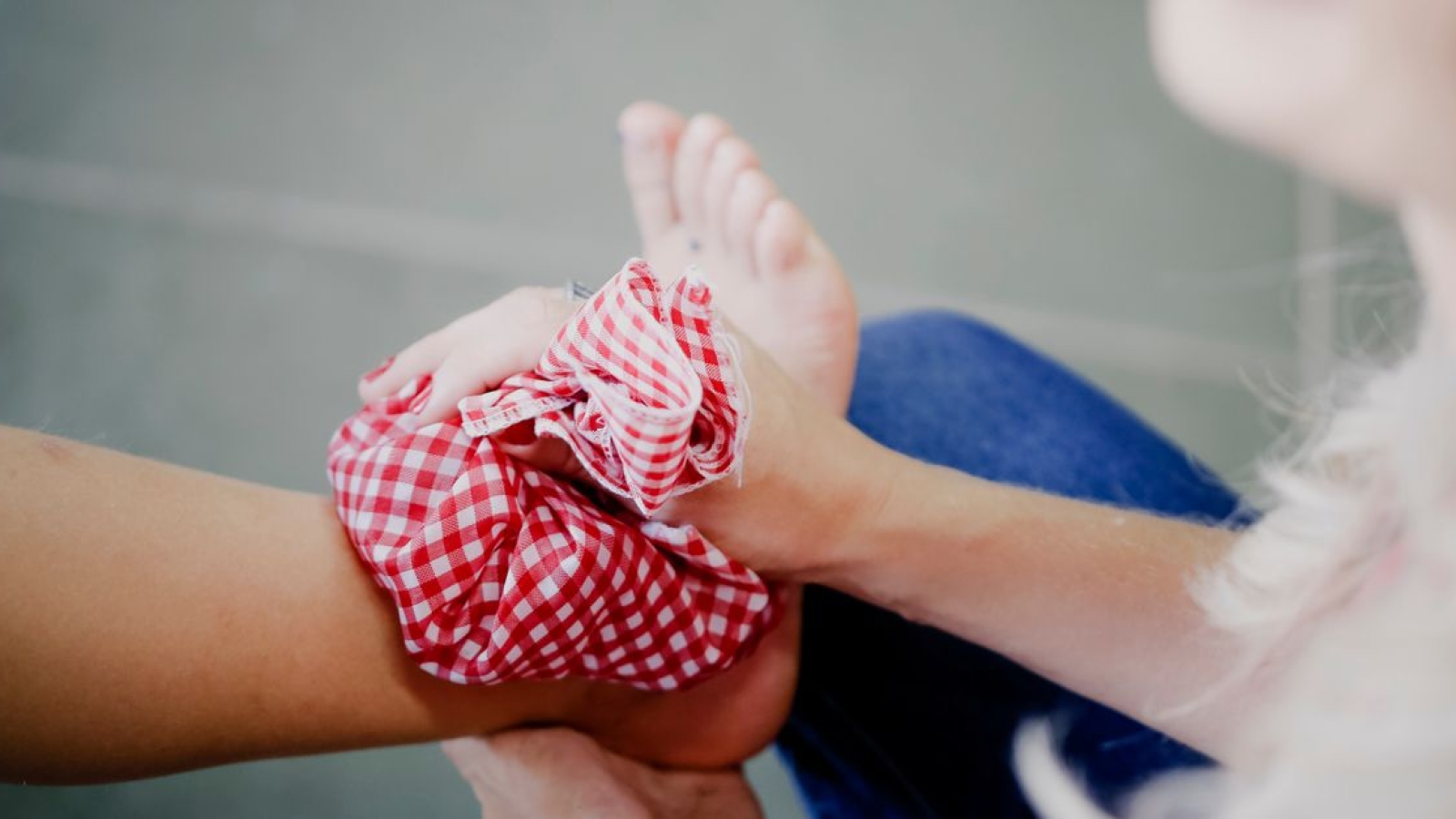
  • Cryotherapy: Increase extensibility and reduce tone of local muscles
  • Ultrasound
  • TENS
  • Deep tissue massage and stretching exercises: Improve extensibility of the muscles who surround the brachial plexus and radial nerve
  • Dry needling: Increase extensibility and reduce tone of local muscles
  • Neural mobilizations:[20]
    • Reduce mechanical extra and intra-neural adhesion
    • Assist the neuromodulation of symptoms
  • Manual therapy[7]: Regain elbow mobility
  • Strengthening[12] and range of motion exercises
  • Stretching exercises:
    • Focus on supinator
    • Passive wrist extensions stretches:
      • Place hand on table and move upper body over wrist
      • Prayer stretch

Post-surgical rehabilitation

  • Commence active range of motion from day 3-5
    • Incorporate stretching of extensors
  • Commence strengthening from week 3-4

Patients can return to light duty work between week 2 and 3 post-operatively, while return to baseline function can take between 6 and 12 weeks.

[21]

Clinical bottom line

The posterior interosseous nerve is the deep branch stemming off the radial nerve. Compression can be caused by trauma, repetitive strain and inflammation. This is then known as posterior interosseous nerve syndrome, which may result in paresis or paralysis of the digital and thumb extensor muscles, resulting in an inability to extend the thumb and fingers at their metacarpophalangeal joints. Conservative management includes splinting, NSAID’s and physiotherapy, and symptoms normally resolve within 3-6 months. Failed conservative management is an indication for surgery, where nerve releases are the most common surgical intervention. Physiotherapy also plays a big part in the post-operative management, and rehabilitation generally lasts between 6 and 12 weeks.

References

  1. ↑ 1.01.11.21.31.4 Vrieling C, Robinson PH, Geertzen JH. ↑ 2.02.1 Chien AJ, Jamadar DA, Jacobson JA, Hayes CW, Louis DS. ↑ 3.03.13.2 Cha J, York B, Tawfik J. Posterior interosseous nerve compression. Eplasty 2014;14.
  2. ↑ 4.04.14.24.3 Bevelaqua AC, Hayter CL, Feinberg JH, Rodeo SA. ↑ 5.05.15.2 Ekstrom RA, Holden K. ↑ Ortho Bullets. PIN Compression Syndrome. Available from: ↑ 7.07.17.2 Quignon R, Marteau E, Penaud A, Corcia P, Laulan J. ↑ 8.08.1 Molina AP, Bour C, Oberlin C, Nzeusseu A, Vanwijck R. ↑ Andreisek G, Crook DW, Burg D, Marincek B, Weishaupt D. ↑ 10.010.110.2 Huisstede BM, Miedema HS, Van Opstal T, De Ronde MT, Kuiper JI, Verhaar JA, Koes BW. ↑ Rosenbaum R. Disputed radial tunnel syndrome. Muscle & Nerve: Official Journal of the American Association of Electrodiagnostic Medicine 1999;22(7):960-7.
  3. ↑ 12.012.112.212.3 Saratsiotis J, Myriokefalitakis E. ↑ 13.013.1 Millender LH, Nalebuff EA, Holdsworth DE. ↑ 14.014.114.2 Kaswan S, Deigni O, Tadisina KK, Totten M, Kraemer BA. Radial tunnel syndrome complicated by lateral epicondylitis in a middle-aged female. Eplasty 2014;14.
  4. ↑ Singh VA, Michael RE, Dinh DB, Bloom S, Cooper M. ↑ Mansuripur PK, Deren ME, Kamal RO. ↑ 17.017.117.217.3 Maffulli N, Maffulli F. ↑ 18.018.1 Chang LW, Gowans JD, Granger CV, Millender LH. ↑ Hashizume H, Nishida K, Nanba Y, Shigeyama Y, Inoue H, Morito Y. ↑ 20.020.1 Molloy J, Neville V, Woods I, Speedy D. ↑ The Student Physical Therapist. Posterior interosseous nerve syndrome. Available from:

    Conteúdos relacionados

Síndrome cruzada mais baixa

Síndrome cruzada mais baixa

Definition

The ‘Unterkreuz syndrome’ is also known as pelvic crossed syndrome, lower crossed syndrome or distal crossed syndrome. The lower crossed syndrome (LCS) is the result of muscle strength imbalances in the lower segment. These imbalances can occur when muscles are constantly shortened or lengthened in relation to each other. The lower crossed syndrome is characterized by specific patterns of muscle weakness and tightness that cross between the dorsal and the ventral sides of the body. In LCS there is overactivity and hence tightness of hip flexors and lumbar extensors. Along with this there is underactivity and weakness of the deep abdominal muscles on the ventral side and of the gluteus maximus and medius on the dorsal side.[1] The hamstrings are frequently found to be tight in this syndrome as well. This imbalance results in an anterior tilt of the pelvis, increased flexion of the hips, and a compensatory hyperlordosis in the lumbar spine.

Crossed syndrome.jpg

                                                                           Figure 1: lower crossed syndrome [2]

Clinically Relevant Anatomy

The pelvic crossed syndrome involves weakness of the trunk muscles: Rectus abdominus, Obliques internus abdominis, Obliques externus abdominis and Transversus abdominis, along with the weakness of the gluteal muscles: Gluteus maximus, gluteus medius and gluteus minimus. These muscles are inhibited and substituted by activation of the superficial muscles.

There is co-existing over activity and tightness of the thoracolumbar extensors: Erector spinae, Multifidus, Quadratus lumborum and Latissimus dorsi; and that of the hip flexors: Iliopsoas and Tensor fasciae latae.

The hamstrings compensate for anterior pelvic tilt or an inhibited gluteus maximus.

Characteristics/Clinical Presentation

This muscle imbalance creates joint dysfunction (ligamentous strain and increased pressure particularly at the L4-L5 and L5-S1 segments, the increased thoracic kyphosis and increased cervical lordosis.[3][4]
There are two known subtypes, A and B, of lower crossed syndrome. The two types are similar and involve the same main muscle imbalance characteristics. For type A the imbalance manifests mainly in the hip, while for type B the imbalance mainly manifests in the lower back. The two subgroups can be distinguished based upon the altered postural alignment and also changed regional myofascial activation patterns. An observation of the lower pole of the thorax and the anterolateral abdominal wall shows whether there are problems with the activity level and balance between the diaphragm and transversus abdominis. Mostly, there is an underactivity of the deep transversus associated with either increased or decreased superficial activity in the obliques and rectus.[3]

Type A: The first subgroup is the posterior pelvic crossed syndrome. In this subgroup there is a domination of the axial extensor.[3] Because the hip flexors are shortened, the pelvis is tilted anteriorly and the hip and knee are in slight flexion. Associated with this is an anterior translation of the thorax because of an increased thoraco-lumbar extensor activity.This gives an expression for the compensatory hyperlordosis of the lumbar spine and hyperkyphosis in the transition from thoracic to lumbar spine. This leads to a decrease in the quality of breathing and of the postural control. Above that the entire thorax will move up, due to the minimal inferior stabilization created by the abdominals. The infra-sternal angle will go up to more than 90° and the postero-inferior thorax will be hyper-stabilized through which it will cause a limited postero-lateral costo-vertebral movement.[3]   

The more anterior and elevated position of the thorax will disturb the stabilization synergies of the Lower Pelvic Unit. The patient will lift the thorax during inspiration which causes an upper chest breathing pattern. This means that the active exhalation will be difficult, because the abdominal activation fails to bring the thorax down and back into the more expiratory caudal (or neutral) position. The abdominal activation is also not sufficient to create the essential intra abdominal pressure. We will notice that the expiratory phase is shortened. This problem arises when the coordination and co-activation between the transverses and the diaphragm is missing. The patient is forced to use the Central Posterior Clinch behavior, which results in an overactivity of the psoas.[3]

Type B: It is also called ‘The Anterior Pelvic Crossed syndrome. In this type the abdominal muscles are too weak and too short. This is associated with a predominant tendency of the axial flexor activity.[3] The compensation is reflected by a minimal hypolordosis of the lumbar spine, a hyperkyphosis of the thoracic spine and protraction of the head. The pelvis is postured more anteriorly and the knees are in hyperextension.[4]

Type A.jpg

                                                                                    Figure 2: type A [2]

Type B.jpg

                                                                                      Figure 3: type B [2]

Examination

Examination for Lower crossed syndrome should follow the same patterns as for examining a patient for Low Back Pain.

Some specific examination points for LCS include the following:

  • OBSERVATION IN ERECT STANDING AND GAIT 

– Position of the pelvis. There is usually an increase of anterior tilt of the pelvis. This can be associated with increased lumbar lordosis.
– Next the shape, size and tone of the tightened/inhibited muscles. (see Definition/Description)

Hip extension – is examined to analyze the hyperextension phase of the hip in gait. Use straight leg lifting.
Hip abduction –the patient with LCS, will combine the abduction with an lateral rotation and a flexion of the hip.
Trunk curl up – is tested to estimate the interplay between usually strong iliopsoas and the abdominal muscles.

Hip abduction.png

                                                                                      Figure 4: Hip Abduction [5]

Trunk curl up.png

                                                                                   Figure 5: Trunk Curl up [5]

  • PASSIVE EXAMINATION : [5]
    -Hip flexors are tested with the patient in a modified piriformis muscle is tested with the patient in a supine position. If the muscle is tight, the end feel is hard and may be associated with pain deep in the buttocks.
    -Quadratus lumborum is difficult to examine. In principle, passive trunk side bending is tested while the patient assumes a side-lying position. The reference point is the level of inferior angle of the scapula. A simpler screening test entails observation of the spinal curve during active lateral flexion of the trunk.
    -Spinal erectors are also difficult to examine. As a screening test, forward bending in a short sit allows observation of the gradual curvature of the spine.
    -Triceps surae are tested by performing passive dorsiflexion of the foot. Normally, the therapist should be able to achieve passive dorsiflexion to 90 degrees.

Physical Therapy Management

The treatment of tightness is not in strengthening as it would further increase tightness and possibly result in more pronounced weakness. A tight muscle should be stretched efficiently. Stretching of tight muscles results in improved strength of inhibited antagonistic muscles, probably mediated via the Sherrington’s law of reciprocal innervation (level of evidence: 2C). [4] [6]

This may involve purely soft tissue approaches. Stretch the specific muscle for a duration of 15 seconds. A five week active stretching program significantly increases active and passive ROM in the lower extremity. [7] (level of evidence: 1B)

Iliopsoas stretch in thomas position

Iliopsoas stretch (and rectus femoris) [8]

The patient is placed in Thomas position. The not-stretched side is maximally flexed to stabilize the pelvis and flatten the lumbar spine. The other leg is normally in flexed position because of the tightness of the iliopsoas. Push this leg into the neutral position (onto the table). Hold this position 15 seconds.

If you want to integrate the rectus femoris into this stretch, bend the knee more than 90° while performing the iliopsoas stretch (level of evidence: 2C).

Self stretching of hip flexors

Erector spinae stretch [9]

The patient lies supine in the fetal position, their knees to their chest with their arms wrapped around their knees. Exhale and stretch. Hold this position for 15 seconds.

The solution for these common patterns is to identify both the shortened and the weakened structures and to set about normalizing their dysfunctional status. This might involve:

  • deactivating trigger points and removing muscular adhesions. Perform myofascial release and trigger-point massage to the gluteus muscles, iliopsoas and tensor fasciae latae (grades of recommendation: B). [10]
  • Laser or ultrasound therapy on the gluteus medius and tensor fasciae latae.
  • Regaining the normal lumbar flexion mobility
  • Core stabilization exercises to strengthen the abdominal muscles.
  • Re-education of posture and body usage. It is necessary to relearn the specific activiation of every element within the Lower Pelvic Unit. This will establish the important fundamental patterns of intra-pevlic control and will also integrate these patterns into basis functional patterns of movement control iniated from the pelvis.
  • Retraining patients with Posterior Pelvic Crossed Syndrome
    It is important to improve the active exhalation, which will bring the thorax caudally on a stable pelvis. It is important to assist the patient, while maintaining the neutral position.. While executing this exercise, it is crucial that the patient breaths down and not up. The patient has to be able to create sufficient intra-abdominal pressure , while maintaining a regular breathing pattern. 
    The patient has to lie down on his back, in supine supported hip flexion to eliminate gravity. The therapist asks the patient to ‘breathe down to the lower placed hand’. It is then important to encourage an active and long exhalation. This gives the patient the sense of the required action. When the correct pattern is mastered it can be progressed into unsupported hip flexion. It is important to push the ribs wide and back, without lifting the thorax. To realize this, the client is asked to push out sideways into the hands of the therapist.[[11], [3]](grades of recommendation: C)

References

  1. ↑ Key J. The Pelvic Crossed Syndromes: A reflection of imbalanced function in the myofascial envelope; a further exploration of Janda’s work. Journal of bodywork and movement therapies. 2010 July;14:299-301
  2. ↑ 2.02.12.2 Janda V. Muscles and motor control in low back pain: Assessment and management. In: Twomey Lt. Physical therapy of the low back. New York, Edinburgh, London: Churchill Livingston, 1987;253-87
  3. ↑ 3.03.13.23.33.43.53.6 Ishida, H., Hirose, R., Watanabe, S., 2012. Comparison of changes in the contraction of the lateral abdominal muscles between the abdominal drawing-in maneuver and breathe held at the maximum expiratory level. Man. Ther. 17 (5), 427- 431. Level of Evidence: 2C
  4. ↑ 4.04.14.2 Chaitow L., DeLany J.W., (2002). Clinical application of neuromuscular techniques: the lower body: Churchill livingstone. (p.26,36)
  5. ↑ 5.05.15.25.3 Liebenson C. Rehabilitation of the Spine: A Practioner’s Manaul. 2nd ed. Philadelphia, PA: Lippincott Williams & Wilkins; 2007.
  6. ↑ Roberts, J., & Wilson, K. (1999). Effect of stretching duration on active and passive range of motion in the lower extremity. Br J Sports Med , 259-263.fckLRLevel of evidence: 1B
  7. ↑ Roberts, J., Wilson, K. (1999). Effect of stretching duration on active and passive range of motion in the lower extremity. Br J Sports Med , 259-263.fckLRLevel of evidence: 1B
  8. ↑ Liebenson, C. (2007). Evaluation of Muscular Imbalance. In Rehabilitation of the Spine: A Practitioner’s Manual (p. 209). Philadelphia: Lippincott Williams & Wilkins.fckLRLevel of evidence: 2C
  9. ↑ Liebenson, C. (2007). Evaluation of Muscular Imbalance. In Rehabilitation of the Spine: A Practitioner’s Manual (p. 209). Philadelphia: Lippincott Williams & Wilkins.fckLRLevel of evidence: 2C
  10. ↑ Simons D.G., Understanding Effective Treatments of Myofascial Trigger Points: Journal of Bodywork and Movement Therapies, 2002, Volume 6, issue 2. fckLRLevel of evidence: 1A
  11. ↑ Key J. (2013), ‘The core’: Understanding it, and retraining its dysfunction, Journal of Bodywork; Movement Therapies 17, p. 541- 559fckLRLevel of evidence: 1A

Conteúdos relacionados

Padrões Internacionais para Classificação Neurológica da Lesão Medular (ISNCSCI)

Padrões Internacionais para Classificação Neurológica da Lesão Medular (ISNCSCI)

The International Standards for Neurological Classification of Spinal Cord Injury (ISNCSCI) or more commonly referred to as the ASIA Impairment Scale (AIS), was developed by the American Spinal Injury Association (ASIA) as a universal classification tool for Spinal Cord Injury based on a standardized sensory and motor assessment, with the most recent revised edition published in 2011. The impairment scale involves both a motor and sensory examination to determine the sensory and motor levels for the right and left side, the overall neurological level of the injury and completeness of the injury i.e. whether the injury is complete or incomplete. [1]

Defined as the most caudal, intact dermatome for both pin prick and light touch sensation.

Defined by the lowest key muscle function that has a grade of at least 3 (on supine testing), providing the key muscle functions represented by segments above that level are judged to be intact (graded as a 5).

This refers to the most caudal segment of the cord with intact sensation and antigravity (3 or more) muscle function strength, provided that there is normal (intact) sensory and motor function rostrally respectively.

A = Complete. No sensory or motor function is preserved in the sacral segments S4-S5.
B = Sensory Incomplete. Sensory but not motor function is preserved below the neurological level and includes the sacral segments S4-S5, AND no motor function is preserved more than three levels below the motor level on either side of the body.
C = Motor Incomplete. Motor function is preserved below the neurological level, and more than half of key muscle functions below the single neurological level of injury have a muscle grade less than 3 (Grades 0-2).
D = Motor Incomplete. Motor function is preserved below the neurological level, and at least half (half or more) of key muscle functions below the NLI have a muscle grade ≥ 3.
E = Normal. If sensation and motor function as tested with the ISNCSCI are graded as normal in all segments, and the patient had prior deficits, then the AIS grade is E. Someone without a SCI does not receive an AIS grade.

ASIA Exam Worksheet

Neurologic Assessment

Sensory Examination

A key point in each of the 28 dermatomes (from C2-S4-5) is tested bilaterally using light touch and pin-prick (sharp-dull discrimination)[1]. Equipment common to clinical settings, such as a cotton wisp and safety pin, may be used. Appreciation of sensation is tested in comparison to sensation on the patient’s cheek. A three-point scale is used for scoring:
0 = absent
1 = altered (impaired or partial appreciation, including hyperesthesia)
2 = normal or intact (similar as on the cheek)
NT = not testable

Deep anal pressure (formerly deep anal sensation): A gentle pressure is applied to the anorectal wall by the examiner’s fnger[1]. Perceived pressure is graded as absent or present.

Motor Examination

Key muscle functions of the myotomes C5-T1 and L2-S1 are tested bilaterally[1]. A six-point scale is used for scoring:
0 = total paralysis
1 = palpable or visible contraction
2 = active movement, full range of motion (ROM) with gravity eliminated
3 = active movement, full ROM against gravity
4 = active movement, full ROM against gravity and moderate resistance in a muscle specific position
5 = (normal) active movement, full ROM against gravity and full resistance in a muscle specific position expected from an otherwise unimpaired person
5* = (normal) active movement, full ROM against gravity and sufficient resistance to be considered normal if identified inhibiting factors (i.e., pain, disuse) were not present
NT = not testable (i.e., due to immobilization, severe pain such that the patient cannot be graded, amputation of limb, or contracture of >50% of the range of motion)

Voluntary anal contraction: The patient is asked to voluntarily contract the external anal sphincter around the examiner’s finger[1]. The contraction is scored as absent or present.

[2]

Determination of Neurological Level of Injury

The neurological level of injury is determined by identifying the most caudal segment of the cord with both intact sensation and normal antigravity muscle function strength. The sensory level refers to the most caudal, intact dermatome for both light touch and pin-prick sensation (score=2). The motor level refers to the most caudal myotome with a key muscle function of at least grade 3. If there is a discrepancy between the most caudal intact section between the four possible levels of right sensory level, left sensory level, right motor level, or left motor level, the neurological level of injury is considered the most rostral segment of these four levels[1].

Zone of Partial Preservation

In complete injuries (AIS A), the zone of partial preservation refers to dermatomes and myotomes caudal to the sensory or motor level that remains partially innervated[1].

Functional Tests

The ISNCSCI measures neurological status in order to describe a patient’s level of impairment. Functional tests are not included in the ISNCSCI examination. However, functional outcomes are necessary for understanding the functional consequences of the level of impairment [3].
Functional tests that the clinician may consider include:

  • 6-minute walk test (6MWT)
  • 10-meter walk test (10MWT)
  • Berg Balance Scale
  • Walking index for spinal cord injury (WISCI II)
  • Spinal cord injury functional ambulation inventory (SCI-FAI)
  • Timed Up and Go Test (TUG)
  • Graded redefined assessment of strength sensibility and prehension (GRASSP)
  • Modified functional reach test (mFRT)
  • Spinal cord independence measure, version III (SCIM-III)
  • Quadriplegia index of function (QIF)

Evidence

Reliability

The ISNCSCI sensory and motor examinations are reliable when conducted by a trained examiner[4]. Formal training in the administration of the ISNCSCI standards has been shown to improve the accuracy of the examiner’s classification[5].

Validity

The ISNCSCI are validated for injury classification[6].

Construct validity of the ASIA motor score as a measure of recovery following SCI and as an outcome measure for clinical trials is greater when upper and lower extremity motor scales are scored independently and not summated together[6].

Resources

References

  1. ↑ 1.01.11.21.31.41.51.6 Burns S, Biering-Sørensen F, Donovan W, Graves D, Jha A, Johansen M, Jones L, Krassioukov A, Kirshblum, Mulcahey MJ, Schmidt Read M, Waring W. International Standards for Neurological Classification of Spinal Cord Injury, Revised 2011. Top Spinal Cord Inj Rehabil 2012;18(1):85-99.
  2. ↑ Kaplan M. Kaplan – ASIA assessment. Available from: ↑ Kalsi-Ryan S, Wilson J, Yang JM, Fehlings MG. Neurological grading in traumatic spinal cord injury. World Neurosurg 2013. http://dx.doi.org/10.1016/j.wneu.2013.01.007 (accessed 13 October 2013).
  3. ↑ Marino R, Jones L, Kirshblum S, Tal J, Dasgupta A. Reliability and repeatability of the motor and sensory examination of the international standards for neurological classification of spinal cord injury. J Spinal Cord Med 2008;31(2)166-170.
  4. ↑ Schuld C, Wiese J, Franz S, Putz C, Stierle I, Smoor I, Weidner N, EMSCI Study Group, Rupp RR. Effect of formal training in scaling, scoring and classification of the international standards for neurological classification of spinal cord injury. Spinal Cord 2013;51(4):282-8.
  5. ↑ 6.06.1 Graves D, Frankiewicz RG, Donovan WH. Construct validity and dimensional structure of the ASIA motor scale. J Spinal Cord Med 2006;29(1):39-45.

Conteúdos relacionados

Escala de Avaliação da Ataxia Cooperativa Internacional

Escala de Avaliação da Ataxia Cooperativa Internacional

Objective

The International Cooperative Ataxia Rating Scale (ICARS) is an outcome measure that was created in 1997 by the Committee of the World Federation of Neurology with the goal of standardizing the quantification of impairment due to cerebellar ataxia. The scale is scored out of 100 with 19 items and 4 subscales of postural and gait disturbances, limb ataxia, dysarthria, and oculomotor disorders. Higher scores indicate higher levels of impairment.

Intended Population

Clients suffering from cerebellar ataxia. The ICARS has been validated for use in patients with focal cerebellar lesions [1] and hereditary spinocerebellar and Friedrich’s ataxia. [2] [3]

Time taken to complete the test – 30 minutes

Method of Use

The International Cooperative Ataxia Rating Scale (ICARS) (Trouillas et al., 1997) is a 100-point semi-quantitative scale. It is divided into four parts, on the basis of the compartmentalization of cerebellar symptoms (Babinski & Tournay, 1913):

ICARS.JPG

Postural and stance disturbances (subscore: /34)
Limb movement disturbances (subscore: /52)
Speech disorders (subscore: /8)
Oculomotor deficits (subscore: /6)

Posture and Gait Score (total of scores A to G)

A. Walking capacities: 10-m test including half-turn, near a wall

0 Normal
1 Almost normal naturally, unable to walk with feet in tandem
2 Walking without support, but abnormal and irregular
3 Walking without support but with considerable staggering; difficulties in half-turn
4 Walking with autonomous support impossible; episodic support of the wall for a 10-m test
5 Walking only possible with one stick
6 Walking only possible with two special sticks or with a stroller
7 Walking only with accompanying person
8 Walking impossible, even with accompanying person (wheelchair)and so on.

PG: 34 Min.: 0 Max.: 100

Grading for ataxia. Balance assessed using posture and gait (PG) subcomponent

Reference

Cerebellar Disorders – A Practical Approach to Diagnosis and Management; Mario Ubaldo Manto, 4 – Clinical scales: 53-68

Evidence

High inter-rater reliability (ICC50.95)

High test–re-test reliability (ICC50.97)

Adequate internal consistency(Cronbach’s a50.94)

Good internal structural validity

Links

References

References will automatically be added here, see adding references tutorial.

  1. ↑ Schoch, B; et al. (Nov 2007). “Reliability and validity of ICARS in focal cerebellar lesions”. Movement Disorders 22 (15): 2162–2169.
  2. ↑ Schmitz-Hubsch, T; et al. (May 2006). “Reliability and validity of the International Cooperative Ataxia Rating Scale: a study in 156 spinocerebellar ataxia patients”. Movement Disorders 21 (5): 699–704
  3. ↑ Storey, E; Tuck, K.; Hester, R.; Hughes, A.; Churchyard, A. (Feb 2004). “Inter-rater reliability of the International Cooperative Ataxia Rating Scale (ICARS)”. Movement Disorders 19 (2): 190–192.

Conteúdos relacionados

Síndrome do Tremor / Ataxia X Frágil

Síndrome do Tremor / Ataxia X Frágil

Original Editor – Carly Wright, Jillian Mayhew & Shannon Huff

Lead Editors

Definition 

Fragile X-associated tremor/ataxia syndrome (FXTAS) is a late onset (> 50 years) neurodegenerative disorder caused by a gene mutation of 55 to 200 repeating CGG (cytosine, guanine, guanine) trinucleotide sequences in the FMR1 gene (fragile X mental retardation 1) [1]. The most common symptoms are action tremor and gait ataxia [2]. Full mutations (greater than 200 CGG repeats) cause fragile X syndrome, which is a common form of mental retardation [3].

Prevalence & Risk Factors

The current estimation is that 1 in 259 females and 1 in 813 males are carriers of the FXTAS premutation gene [3]. The occurrence of female carriers who go on to develop the syndrome is only 4% (very low compared to male carriers), however this rises to 8% in female carriers older than 50 [4]. In addition, the overall presentation of symptoms is less severe in female patients with FXTAS than it is in males [1]. Out of the male carriers of the gene, 17% in their 50s, 38% in their 60s, 47% in their 70s and 75% in their 80s will develop symptoms [4]. The average age of onset is 60.2 (+/- 7.2), however a greater number of CGG repetitions is associated with an earlier age of onset of symptoms [1].

The most significant risk factor for FXTAS is possessing the fragile X (FMR1) gene mutation [1]. Patients with a family history of fragile X or who are known to have the FMR1 premutation carry a high risk of developing symptoms [1]. Males are also at increased risk, as the disease is far less prevalent and less severe in females [1]. Cognitive deficits are not present in all carriers, however age and repeat size are positively correlated with executive function deficits, the predominant cognitive dysfunction [5].

Clinical Presentation

The clinical presentation for FXTAS is non-specific which causes this syndrome to be under-diagnosed [1]. Common symptoms in FXTAS patients are intention tremor and ataxic gait, as well as parkinsonism, neuropathy cognitive decline (executive function deficits and dementia) and psychiatric features [1][3]. Unlike the full mutation, fragile X syndrome, individuals with FXTAS will most commonly have normal intelligence [3]. Some patients (approximately 25%) will have physical deformities and emotional difficulties [3].

Female patients with FXTAS will often have a slightly different presentation than male patients. Premature ovarian failure is a clinical symptom reported in approximately 20% of women with FXTAS [3]. Females are also more likely to develop mental health problems including depression and anxiety as a result of this syndrome [1]. Male carriers, however, are more likely to develop severe motor deficits, social difficulties and obsessive thinking [1].

Cerebellar ataxia and intention tremor are the principal characteristics of FXTAS; these are the most pronounced and often first noticed symptoms, especially in men who developed symptoms over the age of 50 [1][2]. With cerebellar ataxia, patients exhibit a slow gait that involves lurching and difficulty with tandem stance [1]. Intention tremors are the most common type of tremor in FXTAS patients, although sometimes a resting tremor will develop [1]. Other symptoms are variable and can include parkinsonism, autonomic dysfunction, lower limb muscle weakness and peripheral neuropathy [3]. Peripheral neuropathy leads to reduced reflexes and decreased sensation of vibration in the lower limbs [1].

Specifically, the most commonly reported symptoms are: gait difficulties, impaired fine motor skills, writing impairments, muscle weakness, incontinence, and numbness and pain in the lower extremities [3]. FXTAS is also associated with cognitive deficits; in the initial stages patients may experience a reduction in working memory and executive functions [1]. More advanced cognitive deficits are often interpreted as dementia in the later stages [1]. Changes in behavior and personality may also occur [1].

Diagnostic Procedures

FXTAS can be diagnosed using a combination of clinical and radiological (MRI) signs, or by neuropathological analysis of brain tissue [6]. The following table summarizes the common symptoms used for diagnosis of FXTAS [6].

Major Symptoms Minor Symptoms
Radiological Symptoms 1a. MRI: lesions of white matter of the middle cerebral peduncle or MCP sign*
2a.Neuropathology: FXTAS inclusions in brain cells
1b. MRI: lesions of cerebral white matter
2b. MRI: general brain atrophy (moderate to severe)
Clinical Symptoms 3a. Intention tremor
4a. Ataxic gait
3b. Resting tremor (parkinsonism)
4b. Unusual short-term memory problems (ie. rapidly declining)
5b. Decreased executive function

 *The MCP sign is seen in T2 MRIs where high resonance appears at the middle cerebral peduncles [1].

A definite diagnosis of FXTAS requires radiological/neuropathological investigation, and therefore cannot be diagnosed by a physiotherapist using clinical symptoms alone [6]. A diagnosis of FXTAS may be considered definite, probable, or possible based on the following definitions [6].

A patient can be diagnosed with definite FXTAS if the following is present:
• one major radiological symptom (1a or 2a) AND one major clinical symptom (3a or 4a)
• if brain cells contain FXTAS inclusions (2a)

A patient has probable FXTAS if the following is present:
• two major clinical symptoms (3a AND 4a)
…or…
• one minor clinical symptom (3b, 4b or 5b) AND one major radiological symptom (1a or 2a)

A patient has possible FXTAS if the following is present:
• one major clinical sign (3a or 4a) AND one minor radiological symptom (1b or 2b)

Management / Interventions

Medical Management

Treatment of Tremor:
Propanolol (beta-blocker) and Primidone are commonly used to treat essential tremor (ET) and are the most likely contender to treat tremors in FXTAS [4]. Due to the lack of controlled studies on FXTAS, literature about ET treatment is used as a guide.
Botolinum Toxin (BTX) has recently been used on a trial basis with an FXTAS patient who had a disabling arm tremor [4]. The patient experienced significant functional improvement, with the most benefit being 4-6 weeks after injection and lasting on average three months [4]. Further research with controlled trials is required. In controlled trials of BTX being used to treat ET, some cases resulted in a significant decrease in tremor amplitude [4].

Treatment of Ataxia:
Subjective improvements have been observed with the use of carbidopa/levadopa, dopamine agonists and eldepryl in individuals with gait abnormalities and parkinsonism. Amantadine and buspirone have also been successfully used to treat ataxia, although there is no universally effective treatment for cerebellar ataxia [4]. These medications are unfortunately not tolerated well in ataxia patients [4]. Physical therapy however, can improve strength and gait in patients with ataxia and is typically well tolerated [4].

Treatment of Cognitive Deficits:
Cognitive impairment due to FXTAS is treated using dementia pharmaceuticals, traditionally used for Alzheimer’s disease [4]. Aerobic exercise can modify cognitive function in patients with Alzheimer’s disease, which can likely be extrapolated to other neurodegenerative conditions, such as FXTAS [4].

Treatment of Pain:
Neuropathic pain is common, specifically in the lower extremities, for both men and women and fibromyalgia pain is common for women with FXTAS [4]. Antidepressants, antiepileptics and topical analgesics have been effectively used as treatment [4]. Peripheral neuropathic pain in the lower extremities can be reduced by the application of a Liboderm patch [4].

Physical Therapy Management

There is minimal literature published on physical therapy treatment for FXTAS and best practices have yet to be fully researched and defined [7]. Usually, physical therapy is used to treat individualized symptoms and addresses the functional limitations while sustaining, or even improving, fitness levels [7]. Of significant importance is maintaining strength and preventing falls, due to increasing ataxia and parkinsonism traits [7]. FXTAS manifests itself differently in each person affected, meaning therapeutic intervention is mainly at the individual case level; however, there is evidence to support body-weight-supported treadmill training and exercise in general for reducing depression, anxiety and other behavioural issues [7]. Long term improvements from physical therapy, and more specifically, use of body-weight-supported treadmill training, have been observed in walking speed, cadence and stride length, as well as falls reduction [4].

Differential Diagnosis

FXTAS is commonly misdiagnosed as Parkinson’s disease, essential tremor disorder, Alzheimer disease, dementia, or ataxia of unknown etiology. FXTAS has some similarities in symptoms with other neurodegenerative diseases (which are summarized in the table below), however, FXTAS has an FMR1 gene basis. [4]

Fxtas.JPG

Resources

The following website link is for the National Fragile X Foundation which has a page dedicated to FXTAS.
References

  1. ↑ 1.001.011.021.031.041.051.061.071.081.091.101.111.121.131.141.151.161.17 Berry-Kravis E, Abrams L, Coffey S, Hall D, Greco C, Gane L et al. Fragile X-associated tremor/ataxia syndrome: Clinical features, genetics, and testing guidelines. Movement Disorders. 2007;22(14):2018-2030
  2. ↑ 2.02.1 Apartis E, Blancher A, Meissner W, Guyant-Marechal L, Maltete D, De Broucker T et al. FXTAS: New insights and the need for revised diagnostic criteria. Neurology. 2012;79(18):1898-1907.
  3. ↑ 3.03.13.23.33.43.53.63.7 Jacquemont S, Hagerman R, Leehey M, Grigsby J, Zhang L, Brunberg J et al. Fragile X Premutation Tremor/Ataxia Syndrome: Molecular, Clinical, and Neuroimaging Correlates. The American Journal of Human Genetics. 2003;72(4):869-878.
  4. ↑ 4.004.014.024.034.044.054.064.074.084.094.104.114.124.134.144.15 Hagerman R. Treatment of fragile X-associated tremor ataxia syndrome (FXTAS) and related neurological problems. Clinical Interventions in Aging. 2008;Volume 3:251-262
  5. ↑ Seritan A, Kim K, Benjamin I, Seritan I, Hagerman R. Risk Factors for Cognitive Impairment in Fragile X-Associated Tremor/Ataxia Syndrome. Journal of Geriatric Psychiatry and Neurology. 2016;29(6):328-337
  6. ↑ 6.06.16.26.3 [Internet]. 2017 [cited 3 May 2017]. Available from: FXTAS Testing & Diagnosis — National Fragile X Foundation. (n.d.). Retrieved May 03, 2017, from ↑ 7.07.17.27.3 Lieb-Lundell C. Three Faces of Fragile X. Physical Therapy. 2016;96(11):1782-1790

Conteúdos relacionados

Medida de capacidade de pé e tornozelo

Medida de capacidade de pé e tornozelo

Intended Population

The Foot and Ankle Ability Measure (FAAM) is a self-report outcome instrument developed to assess physical function for individuals with foot and ankle related impairments. This self-report outcome instrument is available in English, German, French and Persian. The Foot and Ankle Ability Measure is a 29-item questionnaire divided into two subscales: the Foot and Ankle Ability Measure, 21-item Activities of Daily Living Subscale and the Foot and Ankle Ability Measure, 8-item Sports Subscale. The Sports subscale assesses more difficult tasks that are essential to sport, it is a population-specific subscale designed for athletes[1][2][3].

The FAAM is identical to the FADI, except for an additional 5 items found on the FADI. Four ‘pain-related’ items of the Foot and Ankle Disability Index and the ability of an individual to sleep are deleted. The Sports subscale of the FAAM remains the same as the FADI Sports subscale.

Each item is scored on a 5-point Likert scale (4 to 0) from ‘no difficulty at all’ to ‘unable to do’. Item score totals, which range from 0 to 84 for the ADL subscale and 0 to 32 for the Sports subscale, were transformed to percentage scores. Higher scores represent higher levels of function for each subscale, with 100% representing no dysfunction.

Eechaute et al [4](level 1) concluded that the FADI and FAAM were the most appropriate, patient-assessed instruments to quantify functional disabilities in patients with chronic ankle instability, but reported that there is need for further research of the FAAM in a specific population of patients with CAI[5][6][7].

Method of Use

The ADL and Sports subscales are scored separately.
The response to each item on the ADL subscale is scored from 4 to 0, with 4 being ‘no difficulty’ and 0 being ‘unable to do’. N/A responses are not counted. The score on each of the items are added together to get the item score total. The total number of items with a response is multiplied by 4 to get the highest potential score. If the subject answers all 21 items, the highest potential score is 84. If one item is not answered the highest score is 80, if two are not answered the total highest score is 76, etc. The item score total is divided by the highest potential score. This value is then multiplied by 100 to get a percentage. A higher score represents a higher level of physical function.

The Sports subscale is scored the same as above, 4 being ‘no difficulty at all’ to 0 being ‘unable to do’. The score on each item are added together to get the item score total. The number of items with a response is multiplied by 4 to get the highest potential score. If the subject answers all 8 items the highest potential score is 32, if one item is not answered the highest potential score is 28, if two are not answered the highest potential score is 24, etc. The item score total is divided by the highest potential score. This value is multiplied by 100 to get a percentage. A higher score represents a higher level of physical function.

For the most valid results it is suggested that scores for the FAAM ADL and Sports subscales be generated only when subjects completed 90% or more of the items (19 of 21 for the ADL and 7 of 8 for the Sports subscales respectively)[1].

Evidence

Evidence FAAM.jpg

Evidence for content validity, construct validity, reliability, and responsiveness was obtained among 243 subjects who were separated into a group expected to change and a group expected to remain stable.[6]

Reliability

The stability of a score with repeated measurements over time is defined as test-retest reliability. It is assessed by having an individual complete the instrument 2 or more times during a period when the individual’s condition is expected to remain stable. Intraclass correlation coefficient and SEM values are commonly used to estimate test-retest reliability.

Evidence for reliability was obtained using the group that was expected to remain stable.[4][6]
– ADL subscale: ICC = 0.89; SEM = 2,1 points
– Sport subscale: ICC = 0.87; SEM = 4,5 points

Validity

Content validity

The specific items on the instrument and what they measure determine the evidence that is based on content. The International Classification of Functioning, Disability and Health model can be used to define item content. According to this model, items can potentially measure the domains of (1) body structure and function and (2) activity and participation.’[6]

For the development of the FAAM, the refined version of the FADI, both experts and patients were involved in the final item reduction. [4]

Construct validity


While evidence for content relates directly to what the items measure, evidence for construct validity assesses how the scores on the instrument relate to other measures of the construct.60 A strong relationship between the scores on the instrument and other measures of the same or a related construct provides evidence for convergent validity; little or no relationship between the scores on the instrument and other measures of distinctly different constructs provides evi¬dence for divergent validity.[6]

The ADL and Sport subscales of the FAAM were correlated to the SF-36 physical function subscale and the SF-36 mental function subscale. Strong correlations were found with the SF-36 physical function subscale (r = .84; r = .78), weak correlations were found with the SF-36 mental function subscale (r = .18; r = .11).[4]

  • RobRoy L. Martin [1](level 2) found evidence of validity for the FAAM ADL subscale as an outcome instrument to measure physical function in individuals with diabetes and foot and/or ankle related disorders, but further research is needed for the Sports subscale in individuals with diabetes.
  • Christopher R. Carcia et al [5](level 3) study provides evidence of construct validity for the FAAM. The FAAM may be used to detect self-reported functional deficits in athletes related to Chronic Ankle Instability. Further research is needed.

Responsiveness

Evidence for responsiveness assesses the ability of the instrument to detect changes in an individual’s status over time. The score on an outcome instrument should increase and decrease if the individual’s condition improves and worsens, respectively.[6]

As well the ADL subscale as the Sport subscale of the FAAM were sensitive to significant changes over time. The Guyatt’s responsiveness index for the ADL subscale and the Sport subscale was respectively 2.75 and 1.40. The sport subscale of the FAAM seems to be less responsive than the ADL subscale. The minimal detectable change (MDC) based on a 95% confidence interval was 5.7 and 12.3 points for the ADL and Sports subscales, respectively.[4]

Significantly different change in scores during 4 week in the group expected to change (P[6]

Links

References

  1. ↑ 1.01.11.2 Martin RL, Hutt DM, Wukich DK. Validity of the Foot and Ankle Ability Measure (FAAM) in Diabetes Mellitus. Foot Ankle Int. 2009 Apr;30(4):297-302
  2. ↑ Walmsley et al. The rheumatoid foot: a systematic literature review of patient-reported outcome measures. Journal of Foot and Ankle Research 2010, 3:12.
  3. ↑ RobRoy L. Martin. Evidence of Validity for the Foot and Ankle Ability Measure (FAAM). 2005
  4. ↑ 4.04.14.24.34.4 Eechaute et al. The clinimetric qualities of patient-assessed instruments for measuring chronic ankle instability: A systematic review. BMC Musculoskeletal Disorders 2007, 8:6 doi:10.1186/1471-2474-8-6.
  5. ↑ 5.05.1 Christopher R. Carcia et al. Validity of the Foot and Ankle Ability Measure in Athletes With Chronic Ankle Instability. Journal of Athletic Training 2008;43(2):179–183.
  6. ↑ 6.06.16.26.36.46.56.66.7 RobRoy L. Martin. A Survey of Self-reported Outcome Instruments for the Foot and Ankle. Journal of orthopaedic sports physical therapy. 2007.
  7. ↑ Stephane Borloz et al. Evidence for Validity and Reliability of a French Version of the FAAM. BMC Musculoskeletal Disorders 2011, 12:40doi:10.1186/1471-2474-12-40.
  8. ↑ Thomas G. Mcpoil et al. Clinical Guidelines, Heel Pain – Plantar Fasciitis: Clinical practice guidelines linked to the international classification of functioning, disability, and health from the orthopaedic section of the American physical therapy association. J Orthop Sports Phys Ther. 2008:38(4). Doi:10.2519/jospt.2008.0302.

Conteúdos relacionados

Índice dinâmico de marcha

Índice dinâmico de marcha

Objective

Blender3D NormalWalkCycle.gif

The Dynamic Gait Index (DGI) was developed as a clinical tool to assess gait, balance and fall risk. It evaluates not only usual steady-state walking, but also walking during more challenging tasks.

See the Dynamic Gait Index

Intended Population

Those with poor balance and at risk of falling such as:

Method of Use

8 functional walking tests are performed by the subject and marked out of three according to the lowest category which applies. 24 is the total individual score possible. Scores of 19 or less have been related to increase incidence of falls.

Equipment needed

Box (Shoebox), Cones (2), Stairs, 20′ walkway, 15” wide

Completion Time

15 minutes

Test Protocol [1] [2]

1. Gait level surface _____

Instructions: Walk at your normal speed from here to the next mark (20′)

Grading: Mark the lowest category that applies.

(3) Normal: Walks 20′, no assistive devices, good sped, no evidence for imbalance, normal gait pattern

(2) Mild Impairment: Walks 20′, uses assistive devices, slower speed, mild gait deviations.

(1) Moderate Impairment: Walks 20′, slow speed, abnormal gait pattern, evidence for imbalance.

(0) Severe Impairment: Cannot walk 20′ without assistance, severe gait deviations or imbalance.

2. Change in gait speed _____

Instructions: Begin walking at your normal pace (for 5′), when I tell you “go,” walk as fast as you can (for 5′). When I tell you “slow,” walk as slowly as you can (for 5′).

Grading: Mark the lowest category that applies.

(3) Normal: Able to smoothly change walking speed without loss of balance or gait deviation. Shows a significant difference in walking speeds between normal, fast and slow speeds.

(2) Mild Impairment: Is able to change speed but demonstrates mild gait deviations, or not gait deviations but unable to achieve a significant change in velocity, or uses an assistive device.

(1) Moderate Impairment: Makes only minor adjustments to walking speed, or accomplishes a change in speed with significant gait deviations, or changes speed but has significant gait deviations, or changes speed but loses balance but is able to recover and continue walking.

(0) Severe Impairment: Cannot change speeds, or loses balance and has to reach for wall or be caught.

3. Gait with horizontal head turns _____

Instructions: Begin walking at your normal pace. When I tell you to “look right,” keep walking straight, but turn your head to the right. Keep looking to the right until I tell you, “look left,” then keep walking straight and turn your head to the left. Keep your head to the left until I tell you “look straight,“ then keep walking straight, but return your head to the center.

Grading: Mark the lowest category that applies.

(3) Normal: Performs head turns smoothly with no change in gait.

(2) Mild Impairment: Performs head turns smoothly with slight change in gait velocity, i.e., minor disruption to smooth gait path or uses walking aid.

(1) Moderate Impairment: Performs head turns with moderate change in gait velocity, slows down, staggers but recovers, can continue to walk.

(0) Severe Impairment: Performs task with severe disruption of gait, i.e., staggers outside 15” path, loses balance, stops, reaches for wall.

4. Gait with vertical head turns _____

Instructions: Begin walking at your normal pace. When I tell you to “look up,” keep walking straight, but tip your head up. Keep looking up until I tell you, “look down,” then keep walking straight and tip your head down. Keep your head down until I tell you “look straight,“ then keep walking straight, but return your head to the center.

Grading: Mark the lowest category that applies.

(3) Normal: Performs head turns smoothly with no change in gait.

(2) Mild Impairment: Performs head turns smoothly with slight change in gait velocity, i.e., minor disruption to smooth gait path or uses walking aid.

1) Moderate Impairment: Performs head turns with moderate change in gait velocity, slows down, staggers but recovers, can continue to walk.

(0) Severe Impairment: Performs task with severe disruption of gait, i.e., staggers outside 15” path, loses balance, stops, reaches for wall.

5. Gait and pivot turn _____

Instructions: Begin walking at your normal pace. When I tell you, “turn and stop,” turn as quickly as you can to face the opposite direction and stop.

Grading: Mark the lowest category that applies.

(3) Normal: Pivot turns safely within 3 seconds and stops quickly with no loss of balance.

(2) Mild Impairment: Pivot turns safely in > 3 seconds and stops with no loss of balance.

(1) Moderate Impairment: Turns slowly, requires verbal cueing, requires several small steps to catch balance following turn and stop.

(0) Severe Impairment: Cannot turn safely, requires assistance to turn and stop.

6. Step over obstacle ____

Instructions: Begin walking at your normal speed. When you come to the shoebox, step over it, not around it, and keep walking.

Grading: Mark the lowest category that applies.

(3) Normal: Is able to step over the box without changing gait speed, no evidence of imbalance.

(2) Mild Impairment: Is able to step over box, but must slow down and adjust steps to clear box safely.

(1) Moderate Impairment: Is able to step over box but must stop, then step over. May require verbal cueing.

(0) Severe Impairment: Cannot perform without assistance.

7. Step around obstacles _____

Instructions: Begin walking at normal speed. When you come to the first cone (about 6′ away), walk around the right side of it. When you come to the second cone (6′ past first cone), walk around it to the left.

Grading: Mark the lowest category that applies.

(3) Normal: Is able to walk around cones safely without changing gait speed; no evidence of imbalance.

(2) Mild Impairment: Is able to step around both cones, but must slow down and adjust steps to clear cones.

(1) Moderate Impairment: Is able to clear cones but must significantly slow, speed to accomplish task, or requires verbal cueing.

(0) Severe Impairment: Unable to clear cones, walks into one or both cones, or requires physical assistance.

8. Steps _____

Instructions: Walk up these stairs as you would at home, i.e., using the railing if necessary. At the top, turn around and walk down.

Grading: Mark the lowest category that applies.

(3) Normal: Alternating feet, no rail.

(2) Mild Impairment: Alternating feet, must use rail.

(1) Moderate Impairment: Two feet to a stair, must use rail.

(0) Severe Impairment: Cannot do safely.

TOTAL SCORE: ___ / 24

Scoring

A four-point ordinal scale, ranging from 0-3. “0” indicates the lowest level of function and “3” the highest level of function.

Total Score = 24

Interpretation  22/24 = safe ambulators

Evidence

Dynamic Gait Index, the 4-item Dynamic Gait Index, and the Functional Gait Assessment show sufficient validity, responsiveness, and reliability for assessment of walking function in patients with stroke undergoing rehabilitation, but the Functional Gait Assessment is recommended for its psychometric properties[4].

Reliability

The DGI showed high reliability and showed evidence of concurrent validity with other balance and mobility scales. It is a useful clinical tool for evaluating dynamic balance in ambulatory people with chronic stroke[5].

DGI total scores, administered by using the published instructions, showed moderate interrater reliability with subjects with vestibular disorders. The DGI should be used with caution in this population at this time, because of the lack of strong reliability[6].

The DGI is a reliable functional assessment tool for multiple sclerosis that correlates inversely with timed walk, showing its concurrent validity[7].

Validity

The DGI, although susceptible to ceiling effects, appears to be an appropriate tool for assessing function in healthy older adults[8]

Responsiveness

In stroke: Moderate responsiveness in depicting change at 2 months & 5 months after treatment[9].

In MS: Scores [10].

Miscellaneous

Evaluating a person’s performance on items of the DGI may be useful in identifying gait deviations and in evaluating gait improvements as a result of interventions[11].

Recent Related Research (from Pubmed)

Failed to load RSS feed from http://eutils.ncbi.nlm.nih.gov/entrez/eutils/erss.cgi?rss_guid=187xsalbOHzoR10RvgmLMPbSYW5SxNvA: There was a problem during the HTTP request: 422 Unprocessable Entity

Read 4 Credit

References

  1. ↑ Herdman SJ. Vestibular Rehabilitation. 2nd ed. Philadelphia, PA: F.A.Davis Co; 2000.
  2. ↑ Shumway-Cook A, Woollacott M. Motor Control Theory and Applications, Williams and Wilkins Baltimore, 1995: 323-324.
  3. ↑ AV Verleih. Dynamic Gait Index.AVI. Available from: http://www.youtube.com/watch?v=JtnjfsVnPsY[last accessed 25/05/13]
  4. ↑ Lin JH, Hsu MJ, Hsu HW, Wu HC, Hsieh CL. Psychometric Comparisons of 3 Functional Ambulation Measures for Patients With Stroke. Stroke. 2010 Jul 29; online article ahead of print
  5. ↑ Jonsdottir J, Cattaneo D. Reliability and validity of the dynamic gait index in persons with chronic stroke. Arch Phys Med Rehabil. 2007 Nov;88(11):1410-5.
  6. ↑ Wrisley D, Walker M, Echternach J, Strasnick B. Reliability of the Dynamic Gait Index in people with vestibular disorders. Arch Phys Med Rehabil. 2003;84:1528–1533
  7. ↑ 16McConvey J, Bennett S. Reliability of the Dynamic Gait Index in individuals with multiple sclerosis. Arch Phys Med Rehabil. 2005;86:130–133.
  8. ↑ Herman T, Inbar-Borovsky N, Brozgol M, Giladi N, Hausdorff JM. The Dynamic Gait Index in healthy older adults: the role of stair climbing, fear of falling and gender. Gait Posture. 2009 Feb;29(2):237-41. Epub 2008 Oct 8.
  9. ↑ Lin, J. H., Hsu, M. J., et al. (2010). Psychometric comparisons of 3 functional ambulation measures for patients with stroke. Stroke 41(9): 2021-2025
  10. ↑ Cattaneo, D., Regola, A., et al. (2006). Validity of six balance disorders scales in persons with multiple sclerosis. Disability and Rehabilitation 28(12): 789-795
  11. ↑ Marchetti GF, Whitney SL, Blatt PJ, Morris LO, Vance JM. Temporal and spatial characteristics of gait during performance of the Dynamic Gait Index in people with and people without balance or vestibular disorders. Physical Therapy, 2008 May;88(5):640-51.

Conteúdos relacionados

Síndrome de Down (Trissomia 21)

Síndrome de Down (Trissomia 21)

Introduction

Down Syndrome (DS) is a chromosomal alteration. Chromosomes are structures found in every cell of the body that contain genetic material and are responsible for determining anything ranging from your eye colour to your height. Typically, each cell has 23 pairs of chromosomes, with half coming from each parent [1]. Down syndrome however, occurs when chromosome 21 has a full or partial extra copy in some, or all, of that individual’s cells. This triple copy is sometimes called trisomy 21 [2]. The altered number of chromosomes leads to common physical features in the DS population, such as:

[3]

The following video “Ted Talk” presented by Karen Gaffney, a person with Down Syndrome, explores numerous contemporary thoughts surrounding DS and challenges society’s preconceptions of people with DS.

Epidemiology

DS is the most commonly occurring chromosomal variance noted world-wide [4], with 1 in 700 births resulting in a child with DS [5]. In the UK alone, there are over 41,000 people living with Down Syndrome, and 750 new people born with DS each year [6]. Birth rates are expected to stay the same, but the total population of persons with DS is expected to rise in the coming years. This is mainly due to medical advancements which have increased life expectancy from age 9 in 1929, to 60 years of age today [7]. With this increase in number and age of this population, there will be a larger demand on health services, such as physiotherapy, and increased challenges for families to overcome.

Additionally, persons with DS already report having problems gaining access to health care [8] with the main barrier being a lack of knowledge about available services [9]. Furthermore, parents of persons with DS also commonly express feeling stressed and uncertain about surrounding care of their child and state that they desire more help from physical activity specialists regarding both education and available interventions [10].

Signs and Symptoms

Though there are many similarities across the DS population, there is great variation in the syndrome. There are three types of DS, each with its own set of challenges and individual variation. The three types of DS are Trisomy 21 (95%), Translocation (3%-4%) and Mosaicism (1%)[11]. Further information on the differences between categories can be found [12]. Whichever the type, persons with DS typically have poorer overall health at a young age and exhibit a greater loss of health, mobility, and increased secondary complications as they age when compared to their non-DS counterparts [13][14]. As a result, persons with DS and their families frequently access a range of health services, including physiotherapy. Although DS itself is not a medical condition, and is simply a common variation in the human form, there are many medical conditions that people with DS frequently experience. These include:

Medical conditions.png
  • Learning difficulties  
  • Poor cardiac health  
  • Thyroid dysfunction  
  • Diabetes  
  • Obesity
  • Digestive problems
  • Low bone density
  • Hearing and Vision loss
  • Dementia and Alzheimer’s disease
  • Depression
  • Leukaemia [15][16][17]

Developmental Milestones

From the time a child is born, they are growing and learning. Each person develops at at different pace. However, some skills are expected to be mastered by a specific age. These are called developmental milestones. Milestones can be physical achievements, language related, or social accomplishments. As physiotherapists, we typically focus on motor skills [17].

The ability to move is essential to human life and development. All children begin developing a wide range of movement skills, or motor skills, starting at birth. These motor skills are wide ranging and often broken down into the sub sections below:

DS Motor Skills.png

Motor skills are key for physical function, but also impact cognitive development.

  • Reaching and grasping allows a child to explore the characteristics of objects in his or her physical world.
  • Sitting promotes the use of arms and hands for playing.
  • Walking allows a child to explore the world more effectively than crawling.
  • Independent movement increases opportunities for social interaction which promotes language learning [4][18].

Developmental Milestones in Children with Down Syndrome

Persons with DS will generally achieve all the same basic motor skills necessary for everyday living and personal independence, however it may be at a later age and with less refinement compared to those without DS [19]. Some adjusted milestones for DS are available below:

[20]

For more in depth developmental milestone charts, please see [21], a more indepth description of milestones and a form for recording and tracking progress can be found [18]. While these milestones are generally agreed upon, studies targeting developmental milestones tend to only examine a small number of people. This makes the information less representative of the entire DS population. Researchers also commonly compare people with DS to their non-DS counterparts of the same age. This is an invalid comparison, and it would be more correct to compare children with DS to non-DS individuals of the same mental age. Despite these limitations, the above listed milestones are widely used and considered accurate [22].

Balance and Down Syndrome

It is common for children with DS to be delayed in reaching common milestones such as sitting independently, standing and walking. One of the contributing factors to the delay of these specific milestones is poor balance. It is well known that persons with DS are often considered floppy, clumsy, uncoordinated and have awkward movement patterns due to balance issues. These balance challenges often follow the child into the teen years and sometimes into adulthood [23]. While impaired balance is difficult on its own, it may also impact development of other motor abilities and cognitive development. Being able to maintain balance allows for exploration, social interaction and overall freedom [24].

Factors Contributing to Poor Balance

  • Ligament Laxity: Persons with DS have elastic/loose joints, allowing for a large range of movement. Although this doesn’t sound like a problem it can lead to joints being less stable, difficult to control and may affect balance.
  • Low Muscle Tone: A common symptom of DS is a ‘floppy’ appearance of limbs, with little activity in the muscles at rest, impacting stationary balance. ‘Floppiness’ does improve over time but can influence balance greatly in early years.
  • Slow Reaction Times/Speed of Movement: Persons with DS often are slower to react and move than their non-DS peers. This means that even if the person feels unsteady, it will take a longer time to react to this feeling, and once it is understood, the corrective movement will also be delayed. Both of these aspects make balance challenging.
  • Differences in Brain Size: Persons with DS typically have smaller cerebellums, which is a part of the brain that contributes to the control of balance. The small size impacts its function, limiting balance reflexes, and causing blurry vision when completing tasks at high speed. Other parts of the brain are also smaller, creating issues with voluntary activities, walking technique and coordination.
  • Poor Postural Control: Typically the posture of a person with DS is slouched – hunched over, with a rounded neck. This prevents the head and body from sitting over the pelvis. Posture is impacted by inaccurate messages being sent to the brain from the body’s sensory system. This leaves people with DS less capable of adapting or making anticipatory adjustments to changing environments [24][25][26].

Strength and Down Syndrome

Another contributing factor to delayed milestones and common challenge with DS, is decreased strength.

During childhood, children with DS do not experience the same amount of muscle growth or strength increase as their peers without DS [27]. This is in part due to the decreased amount of physical activity experienced by people with DS, but is also caused for unknown genetic reasons that research is still investigating. Regardless of the reason, persons with DS consistently fall behind in strength categories when compared to their peers without DS, individuals with DS typically exhibiting 40-50% less strength [28].

Factors Affecting Strength

Decreased strength can have a large impact on the lives of persons with DS. Not only can it lead to complication of activities of daily living, making walking up the stairs, getting out of a seat and other seemingly simple tasks, major obstacles, but it can also lead to other problems. Some of these are listed below:

[29]
  • Increased wear and tear on joints
  • Contributes to reduced balance due to weakness in stability muscles
  • Higher risk of falls
  • Elevated level of fatigue
  • Delayed developmental milestones
  • Increased risk of osteoporosis [30]

Reduced Levels of Physical Activity

The research on physical activity levels in people with Down syndrome is conflicting. However, most research does find people with Down syndrome live highly sedentary lives in which they do not achieve the recommended guidelines for physical activity levels [31][32]. The daily recommended levels of physical activity for children is at least 60 minutes of moderate to vigorous intensity activity, and for adults the recommended levels is at least 150 minutes of moderate aerobic activity each week, including at least two strength session in the week [33][34]. Although people with Down syndrome may have decreased capacity for exercise compared to their peers without DS, the guidelines clearly state that children with DS should still meet the recommended guidelines or do as much physical activity as they can manage [35].

Furthermore, as people with DS age, their physical activity levels fall even further behind their peers without DS [31][36][37]. This trend demonstrates that reduced activity levels are a lifelong issue for children with DS that must be addressed.

Barriers to Physical Activity

Most individuals with Down’s Syndrome have to overcome social and environmental barriers to access physical activity. People with DS face many obstacles with the main barriers being lack of money, transportation, access to programs and support from family and carers. It is a common thought that people with DS are too fragile to participate in exercise. [38].

Poor strength and balance are limitations to both cardiovascular and resistance exercise, however, this needs to be addressed as many individuals with DS are now being classed as obese.Individuals with Down syndrome have been found to have substantially higher rates of obesity compared to the general population [39].Often occurring early on in childhood, obesity was found to remain stable from childhood into adulthood, with slight increases after puberty [40]. Obesity is now recognized as a major health risk for people with Down syndrome [41].

The causes of obesity in the Down syndrome population can be divided into physiological causes and behavioural causes. Physiological causes may include conditions such as hypothyroidism, decreased metabolic rate, increased leptin levels (a hormone which helps regulate hunger), short stature and low levels of lean body mass [42]. Behavioural tendencies such as negative thinking and inattention behaviour may become barriers that prevent vital dietary and lifestyle changes to occur [42].

Shelly Obesity Picture.png

Physical inactivity also increases the chance for the development of other health problems such as diabetes, increased blood pressure, dyslipidaemia, early markers of cardiovascular disease, musculoskeletal disorders, breathing difficulties with worsening of sleep apnea and psychological effects including reduced quality of life [33][42].

Aerobic fitness in both youth and adults with Down syndrome is reduced compared to their peers without DS [43][44]. Studies find that adolescents and young adults with DS have comparable aerobic fitness to non DS older adults (60years +) with heart disease [44]. They also have lessened aerobic abilities, reduced muscular strength and reduced bone mineral density levels by 26% compared to their peers without DS [45].

Benefits of Physical Activity

Overall, strong evidence suggests that regular physical activity can lead to numerous health benefits. Participating in physical activity has a positive impact on people’s health. Benefits include improved cardiovascular, metabolic, musculoskeletal and psychosocial health profiles in people with and without DS [46].

Shelly Physical Activity2.png

The fact that many children with DS reach Developmental Milestones later than their peers may be a contributing factor to lower levels of physical activity during infancy.[47] Onset of independent walking in children with Down syndrome occurs roughly 1 year later in comparison to children with typical development [48]. Earlier walking onset has been observed in infants with Down syndrome who performed greater amounts of high intensity activity at 1 year of age [49]. Changes to physical activity levels in infants with Down syndrome has been suggested to encourage motor development, validating the importance of early physiotherapy intervention [47].

Some health benefits of increased physical activity levels in persons with DS are:

[50]
  • Decreased body fat percentage
  • Decreased body weight
  • Improved cardiovascular fitness
  • Improved muscle strength
  • Decreased depression
  • Reduced risk of osteoporosis[51][52][53][54]

In addition to the health benefits listed above, physical activity is important for people with DS because it:

  • Promotes the development of physical and social skills.
  • Establishes a regular routine around being physically active, leading to better habits in the future.
  • Increases life satisfaction.
  • Prevents secondary conditions associated with DS including diabetes, osteoporosis and dementia [55].

From the evidence, it is clear that physical activity is integral to a person with Down syndrome’s health, fitness and wellbeing [35]. If you feel unsure about what kind of activities to encourage your child to take part in, or would like to know what kind of physical activity groups are out there, then hopefully this next section will provide you with some useful information!

Sensation

In addition to the other challenges facing people with DS, they can also experience sensory issues [56]. Being unable to process sensory information from the environment can be both frustrating and challenging, often leading to inappropriate behaviour as a response [57]. As humans, we use sensory information to gain experience, learn and interact with the world. When sensory feedback is limited, it can impact progress in other areas such as motor development [56]. Sensory difficulties can impact a child’s behaviour and the way they interact with people and objects around them [57].

[58]

What is Sensory Integration?

Information from the environment is processed by our brain and can be interpreted by senses such as sound, touch and movement. The brain then organises this information before directing the body’s response; this is known as sensory integration. Typically, we are able to manage all this continuous processing without really having to think about it. People with Down syndrome aren’t always as able to sort through information, and they can quickly become overloaded and sensitive to stimuli. This is called being hyperesponsive and occurs when the brain ‘short-circuits’. Alternatively the person could become immune to stimuli, or hyporesponsive, which occurs where the brain fails to register input [59]. It’s important to understand that people can’t always be neatly categorised into one or the other and crossover does occur.

[60]

Hyperesponsive Behaviour

Most people enjoy a light touch from a loved one, whether a pat on the hand or a hair ruffle, and a positive response is usually expected, particularly from children. However, some people with Down syndrome can register this as dangerous and may scream or pull away. This may seem like an over-reaction to me or you perhaps but is an appropriate response according to the brain of an individual with DS. You could compare it to us walking down a scary street at night where our bodies would be on full alert. If we spent every day in this hightend state of awareness, exploring the environment may become difficult and our reaction to certain scenarios may changes. Without this exploration, it becomes challenging to learn new skills [59].

Hyporesponsive Behaviour

An individual whose brain fails to register input usually bombards the sensory system, typically by constantly touching objects. Pain is not felt in a ‘typical’ manner, by crying or touching the site of pain. Instead the person may bump into the same object over and over. This may also lead to falling repeatedly. If we were in a pitch-black room and told to find a way out, we would call on our other senses – touch and sound for example. People who are hyposensitive are continuously using their brain in this intense way in order to make sense of their environment [59].

Mental Health and Emotional Wellbeing

It is not uncommon for individuals with Down syndrome to experience challenges with emotional behaviours and mental health. Children with Down syndrome may have difficulties with communication skills, problem solving abilities, inattentiveness and hyperactive behaviours. Adolescents may be susceptible to social withdrawal, reduced coping skills, depression, anxiety, obsessive-compulsive behaviours and sleep difficulties. Adults with DS may have similar experiences as adolescents, with further complications of dementia later in life [61]. Listed below are different mental health conditions.

Depression

Adolescents and adults, and sometimes children with Down syndrome may display depressive symptoms such as sadness, severe social withdrawal, or avoidance of activities that were previously enjoyable. These behaviours tend to be associated with an event that may seem like a normal life occurrence, but is perceived as a great stress to someone with Down syndrome. Such events may include the loss of a household pet, a friend or a sibling who moves away, an illness in the family, or the extended absence of a teacher. Individuals with Down syndrome can be particularly sensitive to changes in their environment and if they do not cope appropriately, this may cause significant psychological distress [62]. Challenges may arise including withdrawal from social and physical activities, which may prolong important development in these areas and impact quality of life. There are a variety of treatment options for depression, including counselling, identifying coping methods for stressful events, medications, and participation in exercise and enjoyable activities [63].

Anxiety

Anxiety in a person with Down syndrome may be displayed by behaviours such as restlessness, panic, fidgeting or excessive worrying. Anxiety is often stimulated by transition to a new or unfamiliar situation or environment. For example, going from home to a different environment such as school, a disruption of a daily routine, or anticipation of a new event [64].This may prove to be a challenge when introducing new activities to individuals with DS so it is important to plan ahead and incorporate new activities gradually into the routine.

Routinised and Compulsive-like Behaviours

Children and adults with Down syndrome have a tendency to follow familiar routines that may appear to be repetitive, compulsive or ritualistic [4]. They may require situations to be ‘just right’ or want to participate in the same activities over and over. These behaviours are often performed to avoid feelings of anxiety [65]. It is important to introduce physical and social activities early in life so they become part of an every day routine. This can have a positive impact on achieving developmental milestones.

Hyperactive and Inattentive Behaviours

Children with Down syndrome may appear to be easily distracted, impulsive, frequently restless and they may have difficulty maintaining attention on tasks. This behaviour may persist into adulthood, however it tends to diminish with age [66]. It often causes a barrier to participation in physical activities due to non-compliance and creates the need for increased supervision [38]. There are medications which are said to reduce these behaviours, however they often trigger adverse side effects. It may be beneficial to channel hyperactive energy into participating in activities that the child enjoys, or encourage an activity that provides a calming effect.

Alzheimer’s

Another challenge the brain can face for those with Down syndrome is Alzheimer’s which is the most common cause of dementia. These two terms, Dementia and Alzheimer’s, are commonly used interchangeably but are in fact different diseases. Dementia typically involves symptoms including memory loss, difficulties with thinking, problem-solving and/or language and these occur due to damage to the brain such as that caused by Alzheimer’s. Down syndrome is not the same as Alzheimer’s nor does it guarantee the development of Alzheimer’s! Current estimates state that roughly 50% of people with Down syndrome will develop dementia due to Alzheimer’s as they age and symptoms only begin to show in the individuals 50s or 60s [67].

There has been a lot of research on the link between Down syndrome and Alzheimer’s. Alzheimer’s is a physical disease of the brain caused by build-up of a protein which forms plaques or tangles. As mentioned earlier people with DS have an extra copy of chromosome 21 which carries a certain gene. This gene produces a protein called amyloid precursor protein (APP) which leads to the aforementioned plaques/tangles in many persons with DS [62]. These plaques/tangles can cause a loss of connections between brain cells, leading to a loss in brain tissue. People with Alzheimer’s also have reduced amounts of certain chemicals in their brain which help to transmit signals in the brain, leading to less effective signal transmitting.

Some common symptoms of Alzheimer’s:

  • Short-term memory loss
  • Reduced interest in activities
  • Social withdrawal
  • Confusion and disorientation
  • Increase in wandering
  • Increased problems in unfamiliar places

Physiotherapy Management

Physiotherapy can play a major role in the management of children with DS; through movement and exercise, manual therapy, and education physiotherapists can empower people to take charge of their own health and participate in their treatment. The aim of treatment is to assist people to live as independently as possible [68].

Not everyone with DS requires physiotherapy and as with most things in life, it depends on the individual’s needs. Although there is no standard treatment plan, effective physiotherapy management of Down syndrome typically involves a combination of sensory integration therapy, neurodevelopment treatment, perceptual-motor therapy and traditional strength and conditioning programs [69].

Traditional therapies for conditions involving difficulties with movements can be repetitive and lack variety [70]. People with Down syndrome often have a reduced attention span, which makes engaging in any therapy challenging, especially when dealing with children [71]. By consistently exposing children to different textures, sounds, environments and movements, physiotherapists can make treatment more interesting and inclusive.

Physiotherapists are commonly consulted to educate individuals and their families as well as provide input on health promotion and long-term condition management [72]. As many treatments often require on going maintenance, physiotherapists should encourage family members to support and implement home treatment plans in an attempt to encourage self-management [73]. Due to the variation in all people and across Down syndrome cases, no one physiotherapy intervention can be prescribed. Interventions are based on the individual’s physical and intellectual needs, as well as his or her personal strengths and limitations [16]. Some of the common issues that physiotherapists will address are:

  • Delayed developmental milestones
  • Balance issues
  • Decreased strength
  • Reduced levels of physical activity
  • Issues with sensation
  • Reduced mental health and emotional well-being
  • High chance of Alzheimer’s disease

Choosing the right intervention based on the problems experienced and the individual child is essential to improve the outcome of treatment. Below are some examples of effective interventions for children with Down syndrome. 

Tummy Time

Infancy is the ideal time to start encouraging movement and motor skills. These skills promote interaction between the child and the environment which will improve cognition, language, social skills and independence. Due to the already high risk of developmental delays in infants with DS, this is an especially important area of focus [74].

Tummy Time is a simple physiotherapeutic intervention used for infants with DS. Parents are encouraged to position the child on his tummy in various positions for extended periods of time. Lying in this position has been found to be extremely beneficial, as it affords infants the chance to develop strength, balance and motor skills against gravity. When this technique is practiced, the infant often achieves motor milestones, such as rolling, sitting and crawling, and improved balance earlier in life. Infants who do not experience enough time on their belly have decreased ability to support their own head at 2 months of age and have further delayed developmental milestones [75]. Some of the possible positions are pictured and described below:

[76]

Tummy Time in children without DS has been proven to be a positive contributor to mastering developmental milestones. This intervention has only recently been investigated specifically for children with DS. There are only a few research studies available, but results have been consistently positive. Tummy time should be started as early as possibly in infants with DS. When a child with DS begins this intervention within the first ten weeks of life, levels of motor development similar to that of half the children without DS are experienced. This technique is easily started at birth and can be performed by parents or caregivers. It is the foundation to motor skill mastery in the first year of life and increases balance, strength and skill attainment as the child grows [74].

Further information on tummy time positions is available [77].

Neurodevelopmental Treatment (NDT)

NDT is an approach which focuses on the quality of movement and coordination rather than individual muscle group function [78]. Therefore, NDT is most effective as an early intervention, before poor compensatory patterns of movement become habitual. As physiotherapists, we can use our hands both to prevent abnormal movement patterns and to facilitate more natural ones. This hands-on approach is achieved by the physiotherapist having several ‘key points of control’, including the head, shoulders, trunk and/or pelvis to guide and alter movement [78].  NDT is usually appropriate for people with DS as they often present with limbs and muscles that may not be as well controlled and may be floppier than desired [70].

Although NDT will be different for every individual, a video with some examples of what a more hands-on therapy approach can look like is seen below.

Sensory Integration Therapy (SIT)

People with Down syndrome often struggle to process information from the environment including things like smell, touch and movement. This is known as sensory integration dysfunction. SIT aims to change how the brain interprets the environment through the very aspects in which they struggle, touch, movement and balance [79]. The video below explains these systems and why they are important in everyday life!

SIT involves a wide range of activities and equipment such as weighted vests, brushes, swings, balls, homemade obstacle courses [80] and even game consoles such as the Wii[70]. These items are all used to provide some form of sensory stimulation. This interactive therapy has been shown to increase focus, reduce disruptive behaviours and improve high functioning tasks such as reading, writing and speech [79].

If SIT is identified as a modality it may be beneficial to work alongside an occupational therapist (OT) . OTs are concerned with how people manage to do meaningful activities and can help by providing equipment which ultimately aims to promote independence.

Traditionally seen as more of an ‘OT thing’ understanding how sensory-based issues can impact motor performance can enhance our practice as physiotherapists, particularly when working with children. There is now a physiotherapy special interest group which offers peer support and shared learning for physiotherapists wanting to gain more experience in, and to promote the role of physiotherapy in SIT [81].

Perceptual-Motor Therapy (PMT)

PMT incorporates activities which help to explore balance, coordination and body awareness and is not skills based. So, rather than being taught a certain skill, individuals are provided with an environment in which to explore and determine what their bodies can do [82]. See video below:

Two Wheeled Bicycle Training

Two Wheeled Bicycle riding helps to improve physical activity, is enjoyable and can lead to increased socialization. The skill of bicycle riding can be learned at a young age or later in life [31]. Studies have shown that people with Down Syndrome often have reduced physical activity levels [47], along with reduced sports participation. Assisted two wheeled bicycle riding has been shown to reduce sedentary time and increase time participating in moderate to vigorous activity [51]. As well, this skill has the potential to increase independence and autonomy, whilst helping to diminish their fears surrounding falling from a bike and getting hurt [51]. If you are searching for an activity for your family to do together, that has the potential to improve your child’s quality of life [83], then assisted two wheeled bicycle riding could be a great activity to try out!

Therapeutic Horseback Riding (Hippotherapy)

This is an activity that promotes friendship, fun and progresses confidence with movement skills. Therapeutic horseback riding is a strategy that uses a horse’s motion to promote training of muscle and balance skills required for everyday life activities [84]. While horse riding, the child will experience movements of the trunk, pelvis and hips, similar to those that would take place during normal walking [85]. Adapting to the horse’s rhythmic movements in different directions further enhances muscle contraction, postural control, weight shifting, and planning of movement patterns [86].

The overall benefits of therapeutic horseback riding include advances in balance, muscle strength and coordination, trunk control, postural stability, and weight bearing abilities [84]. Learning new movement strategies through horse riding can also progress skills such as walking, running and jumping [86]. For more information, please see the video below

Treadmill Training

“The key is if we can get them to walk earlier and better then they can explore their environment earlier and when you start to explore, you learn about the world around you” [87]

Infants with typical development learn to walk independently at about 12 months of age. Babies with Down syndrome typically learn to take independent steps at 24-28 months. These are averages, and averages and developmental milestones often feel like a ticking clock to race against. It is important to reassure parents not to worry if their child is is late reaching milestones, and reinforce that each person develops at his or her own pace.

Helping children to walk is of importance as it allows interaction with the world and it is often a stepping stone to the development of other social, motor and cognitive skills. Walking allows children to engage in other enjoyable tasks, and the endurance achieved in doing so allows them to be active for longer periods of time! More skilled walking is less tiring for children and could allow for more energy to take on the rest of the day.

Research carried out in recent years has suggested that regular walking on a treadmill can significantly improve standing and walking ability in children with DS. Put simply, different research groups have used treadmill training, with varying degrees of speed, time length and frequency and concluded that it is a beneficial intervention for development as well as physical activity [87].

Have a look at the children’s development in the video below

What’s great about treadmill training is that physiotherapists can prescribe it to be carried out in a family’s home. This allows parents and carers to include walking practice around the family’s busy schedule and to suit the child. In addition, it promotes their involvement into their child’s development.

Physiotherapy Interventions Developmental Milestones

Physical characteristics of the child with DS such as low muscle tone, loose joints and decreased strength may influence the speed of mastery or alter the form of the developmental milestone. Persons with DS generally naturally overcome these challenges through perseverance [4].

The goal of physiotherapy is not to ‘speed up’ the rate of development. It is simply to facilitate the development of optimal movement patterns. Depending upon capabilities and adaptations made, physical compensations such as pain or inefficient walking patterns may occur. of a physiotherapist is to provide the building blocks to develop a solid physical foundation for movement and exercise that your family member can build on for life.

Building Blocks 3.jpg

Physiotherapy sessions focusing on developmental milestones should be specifically tailored to each child’s current level of development. It is important to observe the child’s abilities and determine what skills should be learned next. As each person is different, skills should be taught in the way the child learns best. It is important that tasks are broken into smaller parts and practiced using different methods based on individual learning styles and physical make up.

Encouraging the Family to Get Involved

It is important to get family members involved with treatment. Practice at home is essential for mastery, and engaging family participation is key. You can teach the family to:

  • Use their child’s interests to encourage new skill development
  • Build on already mastered skills
  • Focus on what their child is willing to learn
  • Practice often
  • Be patient 

Physiotherapy Interventions for Balance

There are a wide range of physiotherapy interventions that can help improve balance. Some of them have been used for many years, while others are still developing and being introduced. Some common traditional physiotherapy interventions to improve balance in persons with DS are:

[88]
  • Stability Exercise (examples available [89])
  • Corrective positioning (examples available [90])
  • Stair climbing
  • Yoga
  • Hydrotherapy [91][24]

Some new emerging physiotherapy interventions being used to improve balance are: 

  • Hippotherapy
  • Treadmill training
  • Two-wheel bicycle training
  • Tummy Time
  • Perceptual-motor therapy
  • Sensory integration training

Encouraging the Family to Get Involved

  1. Practice Makes Perfect: As with everything in life, practice will improve performance. While it often takes more practice to improve performance of balance in a child with DS, it is possible to increase both speed and accuracy of movement.
  2. Encourage Independent Movement: When a person actively initiates a movement, the brain learns how to control the area being moved. This improves coordination and task performance. 
  3. Follow Individual Interests: A child is more likely to eagerly participate if the activity is one that is enjoyed. Try encouraging the family to incorporate balance training into sports and games.
  4. The Earlier the Better: Starting balance practice early in a child’s life will allow for greater amount of learning time and increase muscle strength at a young age.
  5. It’s Never Too Late: Though it is harder to correct learned bad habits, practice at any time is helpful. It is never too late to start. 
  6. Individuals with DS are more commonly visual learners. This means that they learn better by watching others or copying what they can see rather than responding to verbal instruction. Copy cat is a great game to help a family teach their child new tasks [92] 

Physiotherapy Interventions for Strength

Physiotherapy has been quite successful in strength interventions with persons who have DS. There are many techniques that can be used and resources which can be explored. Some of the most common methods to increase strength are:

Method Description
Endurance training Large groups of muscles working at moderate intensity for a more extended period
Weight training Small groups of muscles working at high intensity for a short period of time
Specific muscle training Targeting specific weak muscles[93]

Each of these techniques has been shown to equally increase exercise capacity, health and quality of life in individuals with DS [94]. While these general workout types work well to increase strength, physiotherapists can also offer more specific exercises based on your child’s needs. Below are evolving exercise ideas you can discuss with your physiotherapist or read more about later in the wiki:

  • Tummy time
  • Treadmill training
  • Falls prevention exercises [95]

Encouraging the Family to Get Involved

In order to achieve good results it is important to advise the family on specific exercises, and correct technique. Increasing compliance to exercise will have positive benefits and can help with improving strength and development:

  • Start early: Encourage the family to introduce strength training from a young age, this may help avoid later complications
  • Encourage family members to do exercises with the child.
  • Encourage other types of activities that can build strength such as sports
  • Incorporate strength training into things a child enjoys [96].

Physiotherapy and Physical Activity

Physiotherapists can play a role in encouraging preventative health promotion with their patients who have DS. According to the World Health Organisation [97], the recommended daily physical activity requirements for children is at least 60 minutes of moderate to vigorous intensity daily physical activity. Your physiotherapist can help recommend specific activities suitable for your child and direct you to resources in your area!

Moderate Activity Vigorous Activity:
Aim Increase heart rate and breathing. May cause a light sweat Make the heart and lungs work harder than moderate intensity activity
Example
  • Brisk Walking
  • Active Play
  • Slow Bicycling
  • Water aerobics
  • Slow Dancing [34]
  • Swimming
  • Tennis
  • Running
  • Fast Bicycling
  • Faster Dancing
  • Hiking

Evidence is also growing to support other fun and creative physiotherapy interventions for your child to be physically active including:

  • Treadmill Training
  • Two Wheeled Bicycle Riding
  • Therapeutic Horseback Riding (Hippotherapy)

Structured accessible programs that make adaptations for children with DS have been identified as key to facilitating participation in physical activity [38]. As well, it has been recommended that introducing diverse and interesting physical activity programmes which avoid over complicated tasks, may be more enjoyable for people with DS [98].

Encouraging the Family to Get Involved

One of the most important facilitators identified for improving physical activity participation levels of people with Down syndrome is the support and motivation they receive from their family and carers [38]. Some parents who were interviewed felt their child was more likely to be active when the physical activity was enjoyable and included being with friends or their siblings [37][99]. Introducing physical activities into a child’s routine will increase familiarity and facilitate increased levels of participation [100]. Encouraging the family to keep an activity or exercise log and organising a routine check as well as providing positive feedback, has previously been a suggested as a helpful method to increase motivation towards physical activity participation [99].

Other tips to help encourage children to be physically active include:

  • Choose an activity that the child will enjoy or wants to do.
  • Encourage childhood games that are traditional and active such as hop scotch, hide and seek or obstacle courses.
  • Use simple ways to get children to be more physically active in daily life such as walking to school, taking the stairs instead of the lift or walking the family dog.
  • Keep things simple; running, jumping, dancing are great physical activities to build a child’s fitness and there are no cost requirements! Encourage parents to join in and get fit too!
  • Give your child lots of positive and encouraging feedback. [35].

Physiotherapy Interventions for Sensory Problems

Physiotherapists have recently become more involved in treatment of sensory issues. While this field is still growing, there are a few areas in which physiotherapy has been successful:

  • Advice on desensitisation and calming procedures
  • Provision of sensory specific activities
  • Creation of sensory and behavioural strategies
  • Sensory integration therapy (SIT) – which will be discussed in depth later in the wiki 

Encouraging the Family to Get Involved

Here are some ideas for incorporating therapy into everyday activities:

  • When brushing teeth at night, try using a vibrating toothbrush to increase tolerance of stimuli.
  • Before doing errands, such as taking their child to the hairdressers, try giving them some chewy sweets for the different texture.
  • Encourage children to participate during meal prep or baking; perhaps by mixing ingredients of different textures or carrying pots and pans of different sizes.
  • Encourage the family to involve their child in grocery chores; may allowing the child to push the trolley at the grocery and help with packing and putting food away.
  • Drinking through a straw, a weighted lap blanket or a big squishy seat cushion can all expose children to different sensory experiences while eating.
  • With bathing, encourage parents to try out different brushes, cloths and soaps. Using crazy soap or shaving foam to draw on the wall is an excellent activity. After bath time wrapping a child tightly in a towel and apply pressure is another way to promote sensory activities. A hug works well, if tolerated!
  • Suggest playtime games like the ‘sandwich game’ –  lie the child in between two pillows so they are effectively the sandwich filling and apply pressure on top to their liking. Any home-made obstacle courses involving jumping, crawling, hopping etc. are usually fun and will be beneficial [101].

Another idea is to create a ‘sensory corner’ which can be effective in reducing stress and produce a safe zone for some children. It can provide stimulation for a hyporesponsive person or create a comfortable retreat for a hyperesponsive person. Making a sensory corner is easy! Just block off a corner of a room and use soft furnishings with different textures. For example, use different carpets and pillows. Often a large beanbag or weighted blanket can provide deep pressure that can have a calming effect. Objects like lava lamps, or aquariums may be visually relaxing. Music or a sensory box filled with various objects that differ in texture and weight can be useful. Each person is unique. An example of a sensory room and sensory box are depicted below:

Sensory Games.jpg

Physiotherapy Interventions for Mental Health and Well-being

Physical activity has demonstrated excellent benefits for the mental well-being of individuals with Down syndrome. The benefits include greater life satisfaction, reduced risk of depression, increased self-esteem, and improved social and behavioural skills [54][38]. Any activity that promotes social interaction and friendship will further enhance mental and emotional well-being. Some recommended interventions are:

  • Therapeutic Horseback Riding (Hippotherapy)
  • Two wheeled bicycle training
  • Sensory integration training
  • Perceptual-motor therapy
  • Hydrotherapy
  • Yoga

Encouraging the Family to Get Involved

  • Work with the family to develop a behaviour treatment plan.
  • Encourage the family to take opportunities to interact with others! While most people learn the majority of their social skills in school and work, people with Down syndrome need to “make every contact count”. Whether it is in therapy, school, work or at home, the opportunity to learn is everywhere!
  • Support groups and therapies are a fantastic way of hitting two bases at once; therapy and socialising.
  • Encourage families to develop a routine and stick to it. Try using visual schedules! This method uses pictures or books to help prepare for upcoming events such as beginning a new school year, going to a friend’s party or moving into a new house.
  • Plan for difficult situations. Try using social stories!
  • Where possible encourage the family to promote positive interactions and reduce the negative ones. Make time for fun every day!
  • Explain the importance of encouraging positive behaviours and positive attitudes [4][63][82]

Below is an example of a visual schedule.

Mental Health Pic 1.png

Physiotherapy Interventions and Alzheimer’s

There is no specific physiotherapy treatment for Alzheimer’s, but there are some measures that can be taken to assist in controlling this disease. Physiotherapists can:

  • Promote physical activity to delay the onset of Alzheimer’s
  • Encourage continued activities of daily living to prevent motor changes associated with Alzheimer’s
  • Assist families in planning for caring for their family member with DS and Alzheimer’s
  • Refer families to specialty services for Alzheimer’s [102]

Another aspect in which physiotherapists have recently been involved is Alzheimer’s screening programs for persons with DS. Since Alzheimer’s is so commonly associated with DS, physiotherapists along with other health professionals have begun running yearly screening clinics. These involve simple interviews and routine health checks to identify people at risk of developing Alzheimer’s. This is a great way to keep on top of this possible problem [103]. 

Encouraging the Family to Get Involved

While there is nothing that can be done to prevent the development of Alzheimer’s, the best defence is awareness. Changes in a person’s actions, memory or communication can be signs that something may be wrong.

Encourging yearly DS screening clinics will help people to monitor changes and get an early warning should they be at risk of developing the disease. Encourage the family to read up on Alzheimer’s and create a possible plan in advance may minimize future stress.

Advise clients about the websites for the Other Challenges Associated With Down Syndrome

Reduced Social Interaction

Although not a Physiotherapy issue as such, it’s worth mentioning that the social lives of persons with Down syndrome can be very different from others. Managing many of the physiotherapy issues mentioned above requires time and effort spent in therapy and carrying out home practice. As a result, persons with DS often find themselves meeting and interacting with their peers less often than non-DS persons. This is something to consider as meeting others is important for developing social and life skills.

[104]

Social skills which can differ in people with DS include:

  • Social understanding and empathy
  • Friendship making
  • Play and leisure skills
  • Personal and social independence
  • Socially appropriate behaviour [105]

These are important skills that need to be addressed. A few suggestions to help parents improve these aspects of a child’s life are listed below:

  • Start from an early age and encourage socialisation
  • Encourage independence in all aspects of life
  • Teach social skills in small steps, with consistent messages
  • Sign their child up for sports teams, other activities they enjoy
  • Encourage parents to talk with teachers and monitor their child’s progress
  • Join play groups with other children with and without DS
  • Provide examples of acceptable social behaviour when watching TV or movies [105]

Transition From Child to Adult Services

Becoming an adult can be confusing and difficult for everyone, especially for people with caring needs or intellectual disabilities (ID). When a person with DS gets older they are transferred from children’s services to adult care. The exact age may change depending on the service or area you are located in. This transition is often accompanied by a change in physiotherapist, services available and a disruption to routine. It is often a stressful time and has historically been an issue for service users. In an attempt to correct this, several pieces of legislation have been set out by the UK government.

The Road Ahead Project was commissioned by the Social Care Institute for Excellence (SCIE) in order to explore what information people with DS and their parents might need during the transition [106].The most common pieces of information families wanted to know were:

  1. Parental roles within the transition process including their rights and entitlements
  2. The local situation – support and resources available
  3. The young person’s rights and responsibilities as an adult including information on self-advocacy, empowerment and keeping safe
  4. All possible options available

The Education Act [107] states that at the time of transition, healthcare professionals have several responsibilities;

  • Provision of written advice including details about services likely to be required in the near future once they have left child services
  • Discussion of transfer to adult services with the individual, their family and GP
  • Facilitation of any necessary referrals
  • Attend individual’s annual review meetings from year 9 onwards

Despite numerous government legislations and guidance, research strongly says that there continues to be a marked variation in the arrangements available for the transition from child to adult services [108]. In order to combat this, some further steps have been taken.

Collaboration

  • Increased collaboration between Child and Adolescent Mental Health Services (CAMHS) and adult intellectual disability services.

More Training for Staff

  • Better education for staff in both adult and child services
  • Improved knowledge of legal changes associated with becoming an adult
  • Expanded awareness regarding available referral options

Better Integration Between Services

  • Different services are unique in their structures and philosophies – if there was more integration between services and increased awareness of each other’s role, a more uniform and continuous service could be provided [109].

How Can You Make the Transition Easier?

[110]

While the transition between services relies heavily on geography and physiotherapy services there are a few ways to make this transition easier:

  • Make a plan with the family in advance.
  • Give information on adult services prior to transitioning.
  • Set a date for the transition to occur.
  • Offer to conduct a joint session between new and old services to ease the transition for service providers and the patient and their family.
  • Include the client and their family in the transition decisions [111].

Remember!

Down syndrome can be challenging not only for the individual, but also for the family. It is common for family members of persons with DS to:

Equality Institute. Self Care. 2014. [Picture].
  • Feel increased levels of stress
  • Experience lower levels of well being
  • Exhibit mild depressive symptoms
  • Have decreased confidence in raising their child
  • Think about their child’s social acceptability
  • Worry about their marriage or their other children [112]

Much research has been done on family dynamics and though results are often unclear, recent investigation is revealing that the increased levels of stress and decreased levels of well-being are evident in parents with a child who has DS for a variety of reasons. Demanding parenting roles, concerns over their family member’s social acceptability and decreased confidence in parenting skills are just a few contributors to high stress levels in parents of persons with DS [113].

For this reason, it is important that you remind family members to take time to focus on themselves. Taking time for themselves may improve both their personal mental health and their families’ overall well being. Though these small things may seem insignificant they can have a dramatic effect on how they feel and the cohesiveness of their family unit [114].

References

  1. ↑ National Down Syndrome Society. What is down syndrome. London: NDSS. ↑ Newton R, Marder L, Puri S. Down syndrome: current perspectives. London: Mac Keith Press. ↑ Kami T. Signs of Down Syndrome. 2008. [Picture]. www.womens-health-advice.com/photos/down-syndrome.html (accessed18 March 2018).
  2. ↑ 4.04.14.24.34.4 National Down Syndrome Society. Down syndrome fact sheet. www.ndss.org/wp-content/uploads/2017/08/NDSS-Fact-Sheet-Language-Guide-2015.pdf (accessed 14 March 2018).
  3. ↑ Cipriani G, Danti S, Carlesi C, Fiorino M. Aging with down syndrome: the dual diagnosis: alzheimer’s disease and down syndrome. American Journal of Alzheimer’s Disease and Other Dementias 2018. www.journals.sagepub.com/doi/pdf/10.1177/1533317518761093 (accessed 14 March 2018).
  4. ↑ Carr J, Collins S.. 50 years with down syndrome: a longitudinal study. Journal of Applied Research in Intellectual Disabilities 2018. https://www.ncbi.nlm.nih.gov/pubmed/29498451 (accessed 14 March 2018).
  5. ↑ Zhu J, Hasle H, Correa A, Schendel D, Friedmant J, Olsen J, Ramussen S. Survival among people with down syndrome. Genetics in Medicine 2013;15:64-69. https://www.nature.com/articles/gim201293 (accessed 12 March 2018).
  6. ↑ Allerton L, Emerson E. British adults with chronic health conditions or impairments face significant barriers to accessing health services. Public Health 2012;126:920-927. ↑ National Health Service. Promoting access to healthcare for people with a learning disability. www.jpaget.nhs.uk/media/186386/promoting_access_to_healtcare_for_people_with_learning_disabilities_a_guide_for_frontline_staff.pdf (accessed 14 March 2018).
  7. ↑ Menear, K. Parents perceptions of health and physical activity needs of children with Down syndrome. Down Syndrome Research 2007;12:60-68. https://library.down-syndrome.org/en-gb/research-practice/12/1/parents-perceptions-health-physical-activity-needs-down-syndrome/ (accessed 12 April 2018).
  8. ↑ Pueschel SM, editor. A parent’s guide to Down syndrome: Toward a brighter future. Brookes Pub; 2001.
  9. ↑ Down Syndrome Limerick. Different types of down syndrome. ↑ British Institute of Learning Disabilities. Supporting older people with learning disabilities. https://www.ndti.org.uk/uploads/files/9354_Supporting_Older_People_ST3.pdf (accessed 18 March 2018).
  10. ↑ Cruzado D, Vargas, A. Improving adherence physical activity with a smartphone application based on adults with intellectual disabilities. BMC Public Health. 2013;13:1173. https://bmcpublichealth.biomedcentral.com/articles/10.1186/1471-2458-13-1173 (accessed 11 March 2018).
  11. ↑ Mazeurek D, Wyka J. Down syndrome: genetic and nutritional aspects of accompanying disorders. Roczniki Panstwowego Zakladu Higieny 2015;66:189-194. ↑ 16.016.1 National Human Genome Research Institute. Learning about Down syndrome. ↑ 17.017.1 Sacks B, Buckley S. What do we know about the movement abilities of children with down syndrome. Down Syndrome News and Updates 2003;2:131-141. ↑ 18.018.1 Layton T. Developmental Scale for Children with Down Syndrome. North Carolina: Extraordinary Learning Foundation. www.dsacc.org/downloads/parents/downsyndromedevelopmentalscale.pdf (accessed 20 March 2018). 
  12. ↑ Kim H, Kim S, Kim J, Jeon H, Jung D. Motor and cognitive developmental profiles in children with down syndrome. Annals of Rehabilitation Medicine 2017;41:97-103. ↑ National Down Syndrome Society. Down Syndrome Developmental Milestones. 2009. [Picture]. ↑ Down Syndrome Medical Interest Group. PCHR insert for babies born with Down syndrome. Nottingham: Down Syndrome Medical Interest Group. www.healthforallchildren.com/wp-content/uploads/2013/04/A5-Downs-Instrucs-chartsfull-copy.pdf (accessed 21 March 2018).
  13. ↑ Frank K, Esbensen A. Fine motor and selfcare milestones for individuals with down syndrome using a retrospective chart review. Journal of Theoretical Social Psychology. 2015;89:719-729. ↑ Georgescu M, Cernea M, Balan V. Postural control in down syndrome subjects. The European Proceedings of Social and Behavioural Sciences. www.futureacademy.org.uk/files/images/upload/ICPESK%202015%2035_333.pdf (accessed 17 March 2018).
  14. ↑ 24.024.124.2 Malak R, Kostiukow A, Wasielewska A, Mojs E, Samborski W. Delays in motor development in children with down syndrome. Medical Science Monitor 2015;21:1904-1910. ↑ Costa A. An assessment of optokinetic nystagmus in persons with down syndrome. Experimental Brain Research 2011;8:110-121. ↑ Saied B, Hassan D, Reza B. Postural stability in children with down syndrome. Medicina Sportiva 2014;1:2299-2304. ↑ Cowley P, Ploutz-Snyder L, Baynard T, Heffernan K, Jae S, Hsu S. Physical fitness predicts functional tasks in individuals with Down syndrome. Med Sci Sports Exercise 2010;42:388-393.
  15. ↑ Mercer V, Stemmons V, Cynthia L. Hip abductor and knee extensor muscle strength of children with and without Down’s syndrome. Phys Ther 2001;1318-26.
  16. ↑ Down Syndrome Society. Boy with Down syndrome Flexing. 2017. [Picture].
  17. ↑ Merrick J, Ezra E, Josef B, Endel D, Steinberg D, Wientroub S. Musculoskeletal problems in Down syndrome. Israeli J Pediatr Orthop 2000;9:185-192.
  18. ↑ 31.031.131.2 Esposito PE, MacDonald M, Hornyak JE, Ulrich DA. Physical activity patterns of youth with Down syndrome. Intellectual and developmental disabilities 2012 Apr;50(2):109-19.
  19. ↑ Phillips AC, Holland AJ. Assessment of objectively measured physical activity levels in individuals with intellectual disabilities with and without Down’s syndrome. PLoS One 2011 Dec 21;6(12):e28618.
  20. ↑ 33.033.1 WHO | Physical Activity and Adults [Internet]. Who.int. 2011 [cited 9 April 2018]. Available from: http://www.who.int/dietphysicalactivity/factsheet_adults/en/
  21. ↑ 34.034.1 WHO | Physical activity and young people [Internet]. Who.int. 2011 [cited 9 April 2018]. Available from: http://www.who.int/dietphysicalactivity/factsheet_young_people/en/
  22. ↑ 35.035.135.2 Shields N, Blee F. Physical activity for children with Down syndrome. 2012.
  23. ↑ Shields N, Dodd KJ, Abblitt C. Do children with Down syndrome perform sufficient physical activity to maintain good health? A pilot study. Adapted Physical Activity Quarterly. 2009 Oct;26(4):307-20.
  24. ↑ 37.037.1 Buckley S. Increasing opportunities for physical activity. Down Syndrome Research and Practice 2007;12:18-19.
  25. ↑ 38.038.138.238.338.4 Barr M, Shields N. Identifying the barriers and facilitators to participation in physical activity for children with Down syndrome. Journal of Intellectual Disability Research. 2011 Nov 1;55(11):1020-33.
  26. ↑ Rimmer JH, Yamaki K, Davis BM, Wang E, Vogel LC. Peer reviewed: Obesity and overweight prevalence among adolescents with disabilities. Preventing chronic disease. 2011 Mar;8(2).
  27. ↑ Basil JS, Santoro SL, Martin LJ, Healy KW, Chini BA, Saal HM. Retrospective study of obesity in children with Down syndrome. The Journal of pediatrics. 2016 Jun 1;173:143-8.
  28. ↑ Bull MJ. Health supervision for children with Down syndrome. 2011.
  29. ↑ 42.042.142.2 Artioli T. Understanding Obesity in Down’s Syndrome Children. Journal of Obesity and Metabolism. 2017 1: 101.
  30. ↑ Mendonca GV, Pereira FD, Fernhall BO. Reduced exercise capacity in persons with Down syndrome: cause, effect, and management. Therapeutics and clinical risk management. 2010;6:601.
  31. ↑ 44.044.1 Baynard T, Pitetti KH, Guerra M, Unnithan VB, Fernhall B. Age-related changes in aerobic capacity in individuals with mental retardation: a 20-yr review. Medicine and science in sports and exercise. 2008 Nov;40(11):1984-9.
  32. ↑ Angelopoulou N, Matziari C, Tsimaras A, Sakadamis V, Mandroukas K. Bone mineral density nd muscle strength in young men with mental retardation. Calcified Tissue International 2000;66:176-180.
  33. ↑ Rowland T. Physical activity, fitness, and children. Physical activity and health. 2007:259-70.
  34. ↑ 47.047.147.2 Pitetti H, Baynard T, Agiovlasitis S. Children and adolescents with Down syndrome, physical fitness and physical activity. Journal of Sport and health Science 2013;2:47-57.
  35. ↑ Ulrich BD, Ulrich DA. Spontaneous leg movements of infants with Down syndrome and nondisabled infants. Child development. 1995 Dec 1;66(6):1844-55.
  36. ↑ Lloyd M, Burghardt A, Ulrich DA, Angulo-Barroso R. Physical activity and walking onset in infants with Down syndrome. Adapted Physical Activity Quarterly. 2010 Jan;27(1):1-6.
  37. ↑ LifeSpan .Physical Activity Pyramid. 2015. [Picture].
  38. ↑ 51.051.151.2 Ulrich DA, Burghardt AR, Lloyd M, Tiernan C, Hornyak JE. Physical activity benefits of learning to ride a two-wheel bicycle for children with Down syndrome: a randomized trial. Physical therapy. 2011 Oct 1;91(10):1463-77.
  39. ↑ Rimmer JH, Heller T, Wang E, Valerio I. Improvements in physical fitness in adults with Down syndrome. American Journal on Mental Retardation. 2004 Mar;109(2):165-74.
  40. ↑ Seron BB, Modesto EL, Stanganelli LC, Carvalho EM, Greguol M. Effects of aerobic and resistance training on the cardiorespiratory fitness of young people with Down Syndrome. Revista Brasileira de Cineantropometria & Desempenho Humano. 2017 Aug;19(4):385-94.
  41. ↑ 54.054.1 Heller T, Hsieh K, Rimmer JH. Attitudinal and psychosocial outcomes of a fitness and health education program on adults with Down syndrome. American Journal on Mental Retardation. 2004 Mar;109(2):175-85.
  42. ↑ Shields N. Getting Active: What Does it Mean for Children With Down Syndrome?. 2016.
  43. ↑ 56.056.1 BruniI M.. Fine motor skills for children with Down syndrome. 2nd ed. Bethesda: Woodbine House Inc., 2006.
  44. ↑ 57.057.1 Lashno M. Sensory integration: observations of children with Down syndrome and Autistic spectrum disorders. Disability Solutions 1999;3:31-35.
  45. ↑ Scott R. Do you know me. 2015. [Picture]. https://psychprofessionals.com.au/sensory-processing-problem/ (accessed 12 April 2018).
  46. ↑ 59.059.159.2 Lashno M. Mixed signals: understanding and treating you child’s sensory processing issues. Baltimore: Woodbine House Inc, 2010.
  47. ↑ Smith-Myles B. Seven Senses. 2000. [Picture].↑ Van Germeren-Oosterom H, Fekkes M, Buitendijk S, Mohangoo A, Bruil J, Van Wouwe J. Development, problem behaviour, and quality of life in a population based sample of eight-year-old children with Down syndrome. Plos One 2011;6:7.
  48. ↑ 62.062.1 National Down Syndrome Society. Mental health issues and Down syndrome. https://www.ndss.org/resources/mental-health-issues-syndrome/ (accessed 30 March 2018).
  49. ↑ 63.063.1 National Down Syndrome Association. For families and carers: depression. https://www.downs-syndrome.org.uk/for-families-and-carers/health-and-well-being/depression-2/ (accessed 02 April 2018).
  50. ↑ Evans D, Canavera K, Kleinpeter F, Maccubbin E, Taga K. The fears, phobias and anxieties of children with autism spectrum disorders and Down syndrome: comparisons with developmentally and chronologically age matched children. Child Psychiatry and Human Development 2005;36:3-26.
  51. ↑ Glenn S, Cunningham C, Nananidou A, Prasher V, Glenholmes P. Routinised and compulsive-like behaviours in individuals with Down syndrome. Journal of Intellectual Disability Research 2015;59:1061-1070.
  52. ↑ Selikowitz M. Down syndrome the facts. 2nd ed. Oxford: Oxford University Press, 1997.
  53. ↑ National Institute on Ageing. Alzheimer’s disease in people with Down syndrome. https://www.nia.nih.gov/health/alzheimers-disease-people-down-syndrome accessed 23 March 2018).
  54. ↑ CSP. What is physiotherapy. 2018. www.csp.org.uk/your-health/what-physiotherapy (accessed 14 April 2018).
  55. ↑ Down Syndrome Association. For Families and Carers. ↑ 70.070.170.2 Wuang YP, Chiang CS, Su CY, Wang C. Effectiveness of virtual reality using Wii gaming technology in children with Down syndrome. Res Dev Disabil 2011;32:312-321.
  56. ↑ Lott I, Dierssen M. Cognitive deficits and associated neurological complications in individuals with Down syndrome. Lancet Neurol. 2010;9:623-633.
  57. ↑ CSP. Learning disabilities physiotherapy. Associated of Chartered Physiotherapists for People with Learning Disabilities. www.acppld.csp.org.uk/learning-disabilities-physiotherapy (accessed13 March 2018).
  58. ↑ Middleton J, Kitchen S. Factors affecting the involvement of day centre staff in the delivery of physiotherapy to adults with intellectual disabilities. Journal of Applied Research in Intellectual Disabilities 2008:21:227-235. www.onlinelibrary.wiley.com/doi/10.1111/j.1468-3148.2007.00396.x/epdf (accessed 11 March 2018).
  59. ↑ 74.074.1 Wentz R. The impact of an early and intense prone positioning program in infants with and without Down syndrome. The University of Michigan 2016. ↑ Wentz E. Importance of initiating a tummy time intervention early in infants with down syndrome. Paediatric Physical Therapy 2017;29:68-75. https://journals.lww.com/pedpt/Fulltext/2017/01000/Importance_of_Initiating_a__Tummy_Time_.20.aspx?WT.mc_id=HPxADx20100319xMP (accessed viewed 18 March 2018).
  60. ↑ Wentz E. Tummy Time Positions. 2017. [Picture]. https://journals.lww.com/pedpt/Fulltext/2017/01000/Importance_of_Initiating_a__Tummy_Time_.20.aspx?WT.mc_id=HPxADx20100319xMP (accessed viewed 18 March 2018).
  61. ↑ Durkin A. Tummy Time. www.dsdaytoday.blogspot.co.uk/2011/03/tummy-time.html (accessed 6 April 2018).
  62. ↑ 78.078.1 Velickovic TD, PERAT MV. Basic principles of the neurodevelopmental treatment. Medicina 2005;42:112-120.
  63. ↑ 79.079.1 Smith J, Weaver L, Fristad M. A systematic review of sensory integration training interventions for children with Down syndrome. Sage Journals 2015;5:10-14.
  64. ↑ Zimmer M, Desch L. Sensory integration therapies for children with developmental and behavioural disorders. Paediatrics 2012;129:1186-1189. https://www.ncbi.nlm.nih.gov/pubmed/22641765 (accessed 7 April 2018).
  65. ↑ Sensory integration education. About us. 2013. https://www.sensoryintegration.org.uk/About-us (accessed19 April 2018).
  66. ↑ 82.082.1 Stein D. Behaviour and Down syndrome: a practical guide for parents. Boston: Children’s Hospital Boston, 2002.
  67. ↑ Ringenbach S, Holzapfel G, Mulvey A, Jimenez A, Benson S, Richter M. The effects of assisted cycling therapy and voluntary cycling on reaction time and measures of executive function in adolescents with Down syndrome. JIDR 2016;60:1073-1080.
  68. ↑ 84.084.1 Giagazoglou P, Arabatzi F, Dipla K, Liga, M, Kellis E. Effect of a hippotherapy intervention program on static balance and strength in adolescents with intellectual disabilities. Res Dev Disabil 2012;33:2265-2270.
  69. ↑ Winchester P, Kendall L, Peters H, Sears N, WINKLEY T. The effect of therapeutic horseback riding on gross motor function and gait speed in children who are developmentally delayed.  Physical and Occupational Therapy in Pediatrics 2002;22:37-50.
  70. ↑ 86.086.1 Champagne D, Dugas C. Improving gross motor function and postural control with hippotherapy in children with Down syndrome: case reports. Physiotherapy Theory Practice 2010;26:564-571.
  71. ↑ 87.087.1 Ulrich DA, Ulrich BD, Angulo-Kinzler RM, Yun J. Treadmill training of infants with Down syndrome: evidence-based developmental outcomes. Pediatrics. 2001;108(5):85.
  72. ↑ Clique Images. Yoga for Girls with Down syndrome. 2015. [Picture]. https://www.stocksy.com/1490017/yoga-for-girls-with-down-syndrome (accessed 21 March 2018).
  73. ↑ NHS. Balance Exercises. ↑ Down Syndrome Awareness. Our beautiful son with Down syndrome at Physical Therapy [Video]. 2011. ↑ Gupta S, Rao B, Kumaran S. Effect of strength and balance training in children with down syndrome: a randomized control trial. Clinical Rehabilitation 2011;25:425-432. www.journals.sagepub.com/doi/pdf/10.1177/0269215510382929 (accessed 19 March 2018).
  74. ↑ Welsh TN, Elliot D. The processing speed of visual and verbal movement information by adults with and without Down syndrome. Adapted Physical Activity Quarterly 2001;18:156-167.
  75. ↑ Rimmer J, Heller T, Wang E, Valerio I. Improvements in physical fitness in adults with Down syndrome. Am J Ment Retard. 2004;109:165-174
  76. ↑ Spruit M, Gosselink R, Trooster T. Resistance vs endurance training in patients with COPD and peripheral muscle weakness. Eur Respir J 2002;19:1072-1078.
  77. ↑ Croce R, Pitetti K, Horvat M. Peak Torque, average power, and hamstrings/quadriceps ratios in nondisabled adults and adults with mental retardation. Arch Phys Med Rehabil 1996;77:369-372.
  78. ↑ Lewis C, Fragala-Pinkham M. Effects of aerobic conditioning and strength training on a child with Down syndrome: a case study. Pediatric Physical Therapy 2005;17:30-36.
  79. ↑ World Health Organisation. Global recommendations on physical activity for health. 2017. www.who.int/dietphysicalactivity/factsheet_recommendations/en/ (accessed 28 March 2018).
  80. ↑ Merzbach V, Gordon D. The benefits of exercise to a Down’s Syndrome population [Internet]. Intellectualdisability.info. 2015 [cited 8 April 2018]. Available from: http://www.intellectualdisability.info/physical-health/articles/the-benefits-of-exercise-to-a-downs-syndrome-population
  81. ↑ 99.099.1 Menear K. Parents’ perceptions of health and physical activity needs of children with Down syndrome. Down Syndrome Research and Practice. 2007 Jul 30;12(1):60-8.
  82. ↑ Mahy J, Shields N, Taylor NF, Dodd KJ. Identifying facilitators and barriers to physical activity for adults with Down syndrome. Journal of Intellectual Disability Research. 2010 Sep 1;54(9):795-805.
  83. ↑ Star Institute. Home activities for sensory processing disorders. https://www.spdstar.org/basic/home-activities (accessed 23 March 2018).
  84. ↑ Barnhart R, Connolly B. Agining and Down syndrome: implications for physical therapy. Physical Therapy 2007;87:1399-1406. https://academic.oup.com/ptj/article/87/10/1399/2742283 (accessed 28 March 2018).
  85. ↑ Backer C, Jervis N. A screening programme for adults with Down syndrome. Nursing Times 2007;10. https://www.nursingtimes.net/a-screening-programme-for-adults-with-downs-syndrome/199994.article (accessed 5 April 2018).
  86. ↑ Blue Hippo Group .Plan Ahead. 2015. [Picture].
  87. ↑ 105.0105.1 Social Development Ireland. Social Development. 2017. https://downsyndrome.ie/social-development-behaviour/ (accessed 25 March 2018).
  88. ↑ SCIE. The Road Ahead. https://www.scie.org.uk/publications/tra/index.asp .(accessed 30 March 2018).
  89. ↑ Her Majesty’s Stationery Office. Education Act and associated Code of Practice. London: HMSO, 1993.
  90. ↑ Slopper P, Beecham J, Clarke S, Franklin A, Moran N, Cusworth L. Models of multi-agency services for transition to audit services for disabled young people and those with complex health needs: impacts and cost. University of York: Social policy research unit. https://kar.kent.ac.uk/32459/2/transitions%5B1%5D.pdf (accessed 30 March 2018).
  91. ↑ Singh P, Paul M, Ford T, Kramer T, Weaver T, Mclaren S, Hovish K, Islam Z, Belling R, White S. Process, outcome and experience of transition from child to adult mental healthcare: multiperspective study. The British Journal of Psychiatry 2010;197:305-312.
  92. ↑ Blue Hippo Group .Plan Ahead. 2015. [Picture].
  93. ↑ Viner R. Transition from paediatric to adult care. Bridging the gaps or passing the buck. British Medical Journal 1999;81:3. www.adc.bmj.com/content/81/3/271 (accessed 14 March 2018).
  94. ↑ Cuskelly M, Cram P, Ruper M. Families of children with down syndrome: what we know and what we need to know. Down Syndrome Research and Practice 2008. https://library.down-syndrome.org/en-gb/research-practice/online/2008/families-down-syndrome-what-need/ (accessed 25 March 2018).
  95. ↑ Most D, Fidler D, Booth C, Kelly J. Stress trajectories in mothers of young children with Down syndrome. Journal of Intellectual Disability Research 2006;50:501-514. https://www.ncbi.nlm.nih.gov/pubmed/16774635 (accessed 24 March 2018). 
  96. ↑ Cram P, Warfield M, Shonkoff J, Krauss M. Children with disabilities: A longitudinal study of child development and parent well-being. Monographs of the Society for Research in Child Development 2001;66:266. https://www.ncbi.nlm.nih.gov/pubmed/11677873 (accessed 26 March 2018). 

Conteúdos relacionados

Síndrome do braço morto

Síndrome do braço morto

Definition/Description


“Dead Arm” is characterized by a sudden sharp or ’paralyzing’ pain when the shoulder is moved forcibly into a position of maximum external rotation in elevation or is subjected to a direct blow. The patient is no longer able to perform a throwing movement with the control and the velocity that he achieved before the injury due to pain and numbness. It’s also called recurrent transient subluxation of the shoulder.[1][2] 
The dead arm syndrome is typically associated with anterior instability and a damaged anterior labrum, probably as a result of subluxation of the humeral head. This causes a transient stretch to the brachial plexus during a hard throw.[3] 
The phenomenon is a disorder that can have different causes. Mostly it are problems of the rotator cuff or the labrum. Instability of the shoulder or posterior capsular contracture may be a reason for the development of the dead arm syndrome. In addition, it can also be caused by calcification in the ball and socket joint, bone spurs in the acromion, impingement of the shoulder ligaments, biceps tendonitis, micro-instability, internal impingement and SLAP lesion. Psychological factors can also cause this condition. This syndrome may also occur during throwing, repetitive forceful serving in tennis, or working with the arm in a strained position above shoulder.[1][2][3]  
The symptoms can exacerbate by the loss of the posterior rollback. This leads to anterior translation and results in greater internal impingement posteriorly.[4] 

There are 2 categories of the dead arm syndrome: aware or unaware of subluxation.

Epidemiology / Etiology

The dead arm syndrome is seen most commonly in young athletes (21-30 years) or individuals whose arms have been powerful hyperextended in elevation and external rotation of the shoulder. [1]  
It’s common for people who participate in repetitive throwing sports because the arm is repetitive being turned out backwards as far as possible (external rotation) to create potential energy in the wind up phase prior to the forward acceleration phase. It’s been postulated that the pain is due to an overstretched anterior capsule of the shoulder. Because of this, the ball of the shoulder can shift forward creating an impingement of structures lying in the front of the shoulder joint, which result in pain an the inability to throw. [2]  

Differential Diagnosis 

It’s often misdiagnosed as other shoulder pathology or cervical lesion. There are some factors that differentiate the dead arm syndrome from the other causes of shoulder disability. First it usually appears by young athletic adults (21-30 years). It also has a characteristic history of forceful overextension of the shoulder and there is a positive apprehension test with relocation. [1]

Examination 

There is a positive apprehension test. This test can be carried out when the patient is either in a standing or in a lying position. The shoulder is moved passively into maximum external rotation and in abduction. Then forward pressure is applied to the posterior aspect of the humeral head. The therapist give pressure against the caput humeri to anterior. The test is positive when the patient suddenly becomes apprehensive, complains of pain in the shoulder and has the feeling that the shoulder will come out of the joint considered a positive test. 
In the absence of a strongly positive apprehension test, one should suspect that the shoulder disability is caused by something other than transient subluxation. [1]

Management / Intervention

Treatment includes physical therapy similar to that outlined for shoulder instability and labrum injuries. Surgery may be needed to correct the instability, as well as to repair injuries to the glenoid labrum.[3] 
Once the inflammation and pain have resolved, the patient is subjected to a return to throw program. This takes about 4 weeks.[1] 

Return of full ROM and flexibility is needed before beginning strengthening exercises. These included resisted internal rotation, external rotation, and abduction of the shoulder to strengthen the muscles of the rotator cuff which stabilize the head of the humerus. This program, which is best carried out for three to four months, can decrease the pain and disability.[1][5][3] Rehabilitation of athletes with the dead arm syndrome must include the entire kinetic chain.[1]

Sometimes, it evolves into a full clinical picture of the posteo-superior impingement with a development of a SLAP lesion. Then there is need of a surgical treatment.[6] 
SLAP lesions are repaired through arthroscopy, there are different types of SLAP lesions and the type would determine the repair option.[7]  

Resources

References

  1. ↑ 1.01.11.21.31.41.51.61.7 CR Rowe and B Zarins, Recurrent transient subluxation of the shoulder, J Bone Joint Surg Am. 1981;63:863-872. level of evidence: 2B
  2. ↑ 2.02.12.2 ↑ 3.03.13.23.3 Richard B. Birrer,Bernard A.. Griesemer,Mary B. Cataletto, M.D.Pediatric sports medicine for primary care, 2002, p348
  3. ↑ Donald H. Johnson, M.D, Practical orthopaedic sports medicine & arthroscopy, 2007
  4. ↑ Ho CY, The effectiveness of manual therapy in the management of musculoskeletal disorders of the shoulder: a systematic review, Man Ther. 2009 Oct;14(5):463-74. doi: 10.1016/j.math.2009.03.008. Epub 2009 May 21.
  5. ↑ Kibler WB. The role of the scapula in athletic shoulder function. Am J Sports Med 1998;26:325-337
  6. ↑

Conteúdos relacionados

DPOC (Doença Pulmonar Obstrutiva Crônica)

DPOC (Doença Pulmonar Obstrutiva Crônica)

Introduction

Chronic Obstructive Pulmonary Disease (COPD) is a non-reversible progressive disease that is characterised by symptoms such as shortness of breath, wheezing and a cough that produces a high volume of secretions. It may include Asthma, Emphysema and bronchitis,

Clinically Relevant Anatomy

See this page for information on the anatomy of the lungs

Causes

  • Smoking – The primary risk factor for COPD is chronic tobacco smoking. In the United States, 80 to 90% of cases of COPD are due to smoking.[1]
  • Occupational exposure – Intense and prolonged exposure to workplace dusts found in coal mining, gold mining, and the cotton textile industry and chemicals such as cadmium, isocyanates, and fumes from welding have been implicated in the development of airflow obstruction, even in nonsmokers.[2] Workers who smoke and are exposed to these particles and gases are even more likely to develop COPD. Intense silica dust exposure causes silicosis, a restrictive lung disease distinct from COPD; however, less intense silica dust exposures have been linked to a COPD-like condition.[3] The effect of occupational pollutants on the lungs appears to be substantially less important than the effect of cigarette smoking.[4]
  • Air pollution – Studies in many countries have found that people who live in large cities have a higher rate of COPD compared to people who live in rural areas.[[5] Urban air pollution may be a contributing factor for COPD as it is thought to slow the normal growth of the lungs although the long-term research needed to confirm the link has not been done. In many developing countries indoor air pollution from cooking fire smoke (often using biomass fuels such as wood and animal dung) is a common cause of COPD, especially in women.[6]
  • Genetics – Some factor in addition to heavy smoke exposure is required for a person to develop COPD. This factor is probably a genetic susceptibility. COPD is more common among relatives of COPD patients who smoke than unrelated smokers.[7]] The genetic differences that make some peoples’ lungs susceptible to the effects of tobacco smoke are mostly unknown
  • Autoimmune disease – There is mounting evidence that there may be an autoimmune component to COPD.[8] Many individuals with COPD who have stopped smoking have active inflammation in the lungs.[9]The disease may continue to get worse for many years after stopping smoking due to this ongoing inflammation.[9] This sustained inflammation is thought to be mediated by autoantibodies and autoreactive T cells.[10]

Mechanism of Injury / Pathological Process

It is not fully understood how tobacco smoke and other inhaled particles damage the lungs to cause COPD. The most important processes causing lung damage are:

  • Oxidative stress produced by the high concentrations of free radicals in tobacco smoke.
  • Cytokine release due to inflammation as the body responds to irritant particles such as tobacco smoke in the airway.
  • Tobacco smoke and free radicals impair the activity of antiprotease enzymes such as alpha 1-antitrypsin, allowing protease enzymes to damage the lung.

Clinical Presentation

COPD is a complex interaction between Chronic Bronchitis, Emphysema, and Asthma

Asthma

Asthma is a chronic lung disease which is a very common respiratory condition. It is also known as a reactive airway disease which is inconvenient most of the time but manageable. Asthma is caused by inflammation and constriction of bronchial walls which leads to a series of spasmodic attacks of wheezing and shortness of breath as a result of the hyper-reactivity of smooth muscle in the bronchial walls and in the absence of any other apparent cause. There are various factors such as exposure to cigarette smoke, climate change, physical exertion or emotional stress that causes asthma. It begins during childhood and the disease is commonly triggered by viral infection[11].

Asthma can be diagnosed by the presence of the signs and symptoms. The diagnosis is normally confirmed by presenting a response to a inhaled bronchodialator. Often pulmonary function tests, chest x-rays and blood tests can also be done in order to confirm the diagnosis.

Chronic bronchitis

Lung damage and inflammation in the large airways results in chronic bronchitis. Chronic bronchitis is defined in clinical terms as a cough with sputum production on most days for 3 months of a year, for 2 consecutive years.[12] In the airways of the lung, the hallmark of chronic bronchitis is an increased number (hyperplasia) and increased size (hypertrophy) of the goblet cells and mucous glands of the airway. As a result, there is more mucus than usual in the airways, contributing to the narrowing of the airways and causing a cough with sputum. Microscopically there is infiltration of the airway walls with inflammatory cells. Inflammation is followed by scarring and remodeling that thickens the walls and also results in narrowing of the airways. As chronic bronchitis progresses, there is squamous metaplasia (an abnormal change in the tissue lining the inside of the airway) and fibrosis (further thickening and scarring of the airway wall). The consequence of these changes is a limitation of airflow.[13]

Patients with advanced COPD that have primarily chronic bronchitis rather than emphysema were commonly referred to as “blue bloaters” because of the bluish color of the skin and lips (cyanosis) seen in them.[14] The hypoxia and fluid retention leads to them being called “Blue Bloaters.”

Emphysema

A lateral chest x-ray of a person with emphysema.
Note the barrow chest and flap diaphragm.

Lung damage and inflammation of the alveoli results in emphysema. Emphysema is defined as enlargement of the air spaces distal to the terminal bronchioles, with destruction of their walls.[12] The destruction of air space walls reduces the surface area available for the exchange of oxygen and carbon dioxide during breathing. It also reduces the elasticity of the lung itself, which results in a loss of support for the airways that are embedded in the lung. These airways are more likely to collapse causing further limitation to airflow. The effort made by patients suffering from emphysema during exhalation, causes a pink color in their faces, hence the term commonly used to refer to them, “pink puffers”.

Diagnostic Procedures

Unfortunately there is no single diagnostic test for COPD; diagnosis relies on the presence/absence of symptoms and clinical judgement. The best approach is to undertake a detailed subjective history and physical examination. As COPD is not curable the earlier that it is diagnosed, the earlier treatment can start and that may help to slow down the progression of the disease and the subsequent damage to the lungs.

  • Assessment – A diagnosis of COPD should be considered in patients over the age of 35 who have a risk factor (generally smoking) and who present with exertional breathlessness, chronic cough, regular sputum production, frequent winter ‘bronchitis’ or wheeze.
  • Spirometry – The presence of airflow obstruction should be confirmed by performing post-bronchodilator spirometry. All health professionals involved in the care of people with COPD should have access to spirometry and be competent in the interpretation of the results
  • X-Ray – An x-ray of the chest may show an over-expanded lung (hyperinflation) and can be useful to help exclude other lung diseases.
  • Pulmonary function tests – Complete pulmonary function tests with measurements of lung volumes and gas transfer may also show hyperinflation and can discriminate between COPD with emphysema and COPD without emphysema.
  • Blood tests – A blood sample taken from an artery can be tested for blood gas levels which may show low oxygen levels (hypoxemia) and/or high carbon dioxide levels (respiratory acidosis). A blood sample taken from a vein may show a high blood count (reactive polycythemia), a reaction to long-term hypoxemia.

Outcome Measures

Follow the link Lung Function – Forces Expiratory Volume in 1 second (FEV1)

It is known that COPD lungs lose function quicker and more rapidly than non-COPD lungs. In recognition of this FEV1 is the most important marker to determine severity and treatment in COPD algorithms, with decline of FEV1 over-time as the marker for disease progression[15]. The ratio of FEV1/FVC (Forced vital capacity) as well as the percentage predicted FEV1 is a fixed ratio used in current guidelines to assess the function of lungs.

The strengths of using this measure is that:

  • FEV1 and FVC measurements are highly reproducible
  • Poor lung function if a risk factor for all cause of cardiovascular mortality and poorer health[16]

Limitations being:

  • FEV1 measurements are based on an artificial manoeuvre and do not always correlate with clinically relevant outcomes such as dyspnoea, health status, exercise capacity, or exacerbations[17][18]
  • Patients with similar FEV1 may represent different underlying phenotypes.
  • Reference equations for lung function by European Community for Coal and Steel are disputed and limited in predicting lung function in the general population [19]
  • No minimal important difference (MID) has been established yet. It was suggested that an appropriate range of values for the MID for FEV1 might be 100-140 mL but the MID for FEV1 remains poorly defined for COPD [20]

Lung Volumes

Changes in absolute lung volumes can occur in COPD patients even in the absence of FEV1 changes. Progressive hyperinflation due to airflow limitation and loss of lung elastic recoil not only increases the work required during inspiration but also profoundly decreases the ventilatory reserve and increases the sense of effort and dyspnoea[21]

In terms of measurement static lung hyperinflation and its increase during exercise (dynamic hyperinflation) are measured as elevations of total lung capacity (TLC), functional residual capacity (FRC), residual volume (RV) and as a decrease in inspiratory capacity (IC)[15].

Strengths of lung volumes include:

  • Indices of dynamic hyperinflation correlate better than FEV1 with activity limitation and exertional dyspnoeaand pharmacological and surgical lung volume reduction have been associated with improvements in exercise performance and dyspnoea[22][23]
  • A severely reduced IC/TLC ratio with a threshold value of 25% has been shown to predict mortality in COPD patients[24]

Weaknesses include:

  • Body plethysmography remains the gold standard for the measurement of lung volumes such as TLC, FRC and RV. Spirometrically derived assessments of lung hyperinflation are more difficult to interpret in the absence of simultaneous bodyplethysmographic volume measurements to rule out a concomitant restrictive ventilatory disorder[25]
  • The reproducibility of FRC, IC and RV in absolute values has yet to be demonstrated. Measurement of IC alone is not a reliable marker of lung hyperinflation and does not consistently reflect changes in FRC or TLC[25]
  • The natural course of dynamic hyperinflation in COPD is unknown and seems likely to be highly variable among COPD patients [25]

Exercise Capacity

Includes the Management / Interventions

As COPD is not curable the aim of treatment and interventions are directed at improving quality of life by managing symptoms and exacerbations and slowing down damage to the lungs.

Stopping Smoking

Encouraging patients with COPD to stop smoking is one of the most important components of their management. All COPD patients still smoking, regardless of age, should be encouraged to stop, and offered help to do so, at every opportunity.

Exercise

Exercise prescription is a key component of pulmonary rehabilitation programmes, which are part of the non-pharmacological approach to managing COPD. There is a high level of evidence for the benefits of pulmonary rehabilitation for people with COPD[27] Strength and endurance exercise are endorsed for people with COPD.[28]

Muscles that are required for arm exercise are also involved in movement of the chest wall during respiration and thus the need to breathe often compromises the individual’s ability to undertake daily activities, therefore exercise prescription involving arm exercise needs to be carefully prescribed.[29]

Promote Effective Inhaled Therapy

In people with stable COPD who remain breathless or have exacerbations despite use of short-acting bronchodilators as required, offer the following as maintenance therapy:

  • if forced expiratory volume in 1 second (FEV1)≥50% predicted: either long-acting beta2 agonist (LABA) or long-acting muscarinic antagonist (LAMA)
  • if FEV1

Offer LAMA in addition to LABA + ICS to people with COPD who remain breathless or have exacerbations despite taking LABA + ICS, irrespective of their FEV1.

Provide Pulmonary Rehabilitation

Pulmonary rehabilitation should be made available to all appropriate people with COPD including those who have had a recent hospitalisation for an acute exacerbation.

Use Non-Invasive Ventilation

Non-invasive ventilation (NIV) should be used as the treatment of choice for persistent hypercapnicventilatory failure during exacerbations not responding to medical therapy. It should be delivered by staff trained in its application, experienced in its use and aware of its limitations. When patients are started on NIV, there should be a clear plan covering what to do in the event of deterioration and ceilings of therapy should be agreed.

Manage Exacerbations

The frequency of exacerbations should be reduced by appropriate use of inhaled corticosteroids and bronchodilators, and vaccinations.

The impact of exacerbations should be minimised by:

  • giving self-management advice on responding promptly to the symptoms of an exacerbation
  • starting appropriate treatment with oral steroids and/or antibiotics
  • use of non-invasive ventilation when indicated
  • use of hospital-at-home or assisted-discharge schemes

Ensure Multidisciplinary Working

COPD care should be delivered by a multidisciplinary team.

Managing Symptoms and Conditions in Stable COPD

Breathlessness and Exacerbations

  • Manage breathlessness and exercise limitation with inhaled therapy
  • For exacerbations or persistent breathlessness:
    • use long-acting bronchodilators or LABA + ICS
    • consider adding theophylline if still symptomatic
  • Offer pulmonary rehabilitation to all suitable people
  • Refer patients who are breathless, have a single large bulla on a CT scan and an FEV1 less than 50% predicted for consideration of bullectomy
  • Refer people with severe COPD for consideration of lung volume reduction surgery if they remain breathless with marked restrictions of their activities of daily living, despite maximal medical therapy (including rehabilitation), and meet all of the following:
    • FEV1 greater than 20% predicted
    • PaCO2 less than 7.3 kPa
    • upper lobe predominant emphysema
    • TLCO greater than 20% predicted
  • Consider referring people with severe COPD for assessment for lung transplantation if they remain breathless with marked restrictions of their activities of daily living despite maximal medical therapy. Considerations include:
    • age
    • FEV1
    • PaCO2
    • homogeneously distributed emphysema on CT scan
    • elevated pulmonary artery pressures with progressive deterioration
    • comorbidities
    • local surgical protocols

Frequent Exacerbations

  • Optimise inhaled therapy
  • Offer vaccinations and prophylaxis
  • Give self-management advice
  • Consider osteoporosis prophylaxis for people requiring frequent oral corticosteroids

Cor Pulmonale

  • Consider in people who have peripheral edema, a raised venous pressure, a systolic parasternal heave, a loud pulmonary second heart sound
  • Exclude other causes of peripheral edema
  • Perform pulse oximetry, ECG and echocardiogram if features of cor pulmonale
  • Assess need for LTOT
  • Treat edema with diuretic
  • Angiotensin-converting enzyme inhibitors, calcium channel blockers, alpha-blockers are not recommended
  • Digoxin may be used where there is atrial fibrillation

Respiratory Failure

  • Assess for appropriate oxygen
  • Consider referral for assessment for long-term domiciliary NIV therapy

Abnormal BMI

  • Refer for dietetic advice
  • Offer nutritional supplements if the BMI is low
  • Pay attention to weight changes in older patients (especially>3 kg)

Chronic Productive Cough

  • Consider mucolytic therapy

Anxiety and Depression

Alpha-1 Antitrypsin Deficiency

  • Offer referral to a specialist centre to discuss the clinical management of this condition
  • Alpha-1 antitrypsin replacement therapy is not recommended

Palliative Setting

  • Opioids should be used when appropriate for the palliation of breathlessness in people with end-stage COPD unresponsive to other medical therapy
  • Use benzodiazepines, tricyclic antidepressants, major tranquillisers and oxygen to treat breathlessness
  • Provide access to multidisciplinary palliative care teams and hospices

Resources

  • KNGF guidelines for physical therapy in patients with chronic obstructive pulmonary disease

Videos

References

  1. ↑ Young RP, Hopkins RJ, Christmas T, Black PN, Metcalf P, Gamble GD (August 2009). “COPD prevalence is increased in lung cancer, independent of age, sex and smoking history”. Eur. Respir. J. 34 (2): 380–6
  2. ↑ Devereux, Graham (May 2006). “ABC of chronic obstructive pulmonary disease. Definition, epidemiology, and risk factors”. BMJ 332 (7550): 1142–4
  3. ↑ Hnizdo E, Vallyathan V (April 2003). “Chronic obstructive pulmonary disease due to occupational exposure to silica dust: a review of epidemiological and pathological evidence”. Occup Environ Med 60 (4): 237–43
  4. ↑ Loscalzo, Joseph; Fauci, Anthony S.; Braunwald, Eugene; Dennis L. Kasper; Hauser, Stephen L; Longo, Dan L. (2008). Harrison’s Principles of Internal Medicine (17th ed.). McGraw-Hill Professional
  5. ↑ Halbert RJ, Natoli JL, Gano A, Badamgarav E, Buist AS, Mannino DM (September 2006). “Global burden of COPD: systematic review and meta-analysis”. Eur. Respir. J. 28 (3): 523–32
  6. ↑ Kennedy SM, Chambers R, Du W, Dimich-Ward H (December 2007). “Environmental and occupational exposures: do they affect chronic obstructive pulmonary disease differently in women and men?”. Proceedings of the American Thoracic Society 4 (8): 692–4.
  7. ↑ ilverman EK, Chapman HA, Drazen JM, et al. (June 1998). “Genetic epidemiology of severe, early-onset chronic obstructive pulmonary disease. Risk to relatives for airflow obstruction and chronic bronchitis”. Am. J. Respir. Crit. Care Med. 157 (6 Pt 1): 1770–8
  8. ↑ Agustí A, MacNee W, Donaldson K, Cosio M. (2003). “Hypothesis: does COPD have an autoimmune component?”. Thorax 58 (10): 832–4
  9. ↑ 9.09.1 Rutgers, Steven R.; Postma, Dirkje S.; Ten Hacken, Nick H. .T.; Kauffman, Henk F.;van der Mark,Thomas W; Koeter, Gerard H.; Timens, Wim (2000). “Ongoing airway inflammation in patients with COPD who do not currently smoke”. Thorax 55 (1): 12–18.
  10. ↑ Feghali-Bostwick CA, Gadgil AS, Otterbein LE, et al. (January 2008). “Autoantibodies in patients with chronic obstructive pulmonary disease”. Am. J. Respir. Crit. Care Med. 177 (2): 156–63. doi:10.1164/rccm.200701-014OC
  11. ↑ http://www.atsjournals.org/doi/abs/10.1164/rccm.200809-1512OC
  12. ↑ 12.012.1 Longmore, J. M.; Murray Longmore; Wilkinson, Ian; Supraj R. Rajagopalan (2004). Oxford handbook of clinical medicine. Oxford [Oxfordshire]: Oxford University Press. pp. 188–9. ISBN 0-19-852558-3
  13. ↑ Kumar P, Clark M (2005). Clinical Medicine (6th ed.). Elsevier Saunders. pp. 900–1. ISBN 0702027634
  14. ↑ Chung C, Delaney J, Hodgins R (2008). “Respirology”. in Somogyi, Ron; Colman, Rebecca. The Toronto notes 2008: a comprehensive medical reference and review for the Medical Council of Canada Qualifying Exam – Part 1 and the United States Medical Licensing Exam – Step 2. Toronto: Toronto Notes for Medical Students. p. R9. ISBN 0-9685928-8-0
  15. ↑ 15.015.1 Glaab T. Vogelmeier C and Buhl R. Outcome measures in chronic obstructive pulmonary disease (COPD): strengths and limitations. Respiratory Research. 2010:11:79
  16. ↑ Sin DD, Wu L, Man SF: The relationship between reduced lung function and cardiovascular mortality: a population-based study and a systematic review of the literature. Chest 2005, 127:1952-1959
  17. ↑ Cazzola M, MacNee W, Martinez FJ, Rabe KF, Franciosi LG, Barnes PJ, Brusasco V, Burge PS, Calverley PMA, Celli BR, Jones PW, Mahler DA, Make B, Miravitlles M, Page CP, Palange P, Parr D, Pistolesi M, Rennard SI, Rutten-van Mölken MP, Stockley R, Sullivan SD, Wedzicha JA, Wouters EF, American Thoracic Society/European Respiratory Society Task Force on outcomes of COPD: Outcomes for COPD pharmacological trials: from lung function to biomarkers. Eur Respir J 2008, 31:416-469
  18. ↑ 11.Wise RA: The value of forced expiratory volume in 1 second decline in the assessment of chronic obstructive pulmonary disease progression. Am J Med 2006, 119:4-11
  19. ↑ 12.Pellegrino R, Viegi G, Brusasco V, Crapo RO, Burgos F, Casaburi R, Coates A, van der Grinten CPM, Gustafsson P, Hankinson J, Jensen R, Johnson DC, MacIntyre N, McKay R, Miller MR, Navajas D, Pedersen OF, Wanger J: Interpretative strategies for lung function tests. Eur Respir J 2005, 26:948-968
  20. ↑ 4.Cazzola M, MacNee W, Martinez FJ, Rabe KF, Franciosi LG, Barnes PJ, Brusasco V, Burge PS, Calverley PMA, Celli BR, Jones PW, Mahler DA, Make B, Miravitlles M, Page CP, Palange P, Parr D, Pistolesi M, Rennard SI, Rutten-van Mölken MP, Stockley R, Sullivan SD, Wedzicha JA, Wouters EF, American Thoracic Society/European Respiratory Society Task Force on outcomes of COPD: Outcomes for COPD pharmacological trials: from lung function to biomarkers. Eur Respir J 2008, 31:416-469
  21. ↑ O’Donnell DE, Laveneziana P: Physiology and consequences of lung hyperinflation in COPD. Eur Respir Rev 2006, 15:61-67
  22. ↑ 17.O’Donnell DE: Is sustained pharmacologic lung volume reduction now possible in COPD? Chest 2006, 129:501-503
  23. ↑ 18.Criner GJ, Belt P, Sternberg AL, Mosenifar Z, Make BJ, Utz JP, Sciurba F: National Emphysema Treatment Trial Research Group. Effects of lung volume reduction surgery on gas exchange and breathing pattern during maximum exercise. Chest 2009, 135:1268-79
  24. ↑ 19.Casanova C, Cote C, de Torres JP, Aguirre-Jaime A, Marin JM, Pinto-Plata V, Celli BR: Inspiratory-to-total lung capacity predicts mortality in patients with chronic obstructive pulmonary disease. Am J Respir Crit Care Med 2005, 171:591-597
  25. ↑ 25.025.125.2 O’Donnell DE, Laveneziana P: Physiology and consequences of lung hyperinflation in COPD. Eur Respir Rev 2006, 15:61-67
  26. ↑ Burke Rehabilitation. COPD Treatments & Rehab: Upper Body Exercises. Available from: http://www.youtube.com/watch?v=VR7QnSnHmBU[last accessed 13/02/15]
  27. ↑ Roisin RR, Rabe KF, Anzueto A, et al. Global strategy for the diagnosis management, and prevention of chronic obstructive pulmonary disease. Bethesda, MD: Global Initiative for Chronic Obstructive Lung Disease, 2008; 1–91.
  28. ↑ Skinner, Margot. Strength and endurance exercise endorsed for people with COPD. Physical Therapy Reviews, Volume 14, Number 6, December 2009 , pp. 418-418(1)
  29. ↑ Ennis S, Alison J, McKeough Z. The effects of arm endurance and strength training on arm exercise capacity in people with chronic obstructive pulmonary disease. Phys Ther Rev 2009;14(4):226–39.
  30. ↑ SMACC. Non-Invasive Ventilation. Available from: http://www.youtube.com/watch?v=QQZvhkBWBgQ [last accessed 13/02/15]
  31. ↑ National Institute for Health and Clinical Excellence. Chronic obstructive pulmonary disease: Management of chronic obstructive pulmonary disease in adults in primary and secondary care. Available from http://guidance.nice.org.uk/CG91 [last accessed 2/8/10]

Conteúdos relacionados